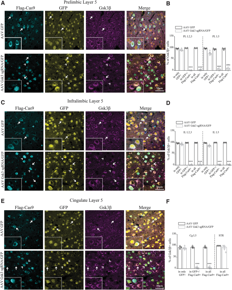
FIG. 2.
Knockout of GSK3β in D2 neurons of the medial prefrontal cortex (mPFC). (A) Immunofluorescent staining for Flag-Cas9 and Gsk3β in prelimbic (PL) layer 5 of virus-injected D2Cre/LSL-Flag-Cas9 mice. Insets show the zoomed picture. (B) Quantification of the percentage of Gsk3β-expressing cells (PL L2/3 AAV GFP condition: in only GFP 96.8 ± 1.7, in GFP+/Flag-Cas9 + 99.2 ± 0.75, in all Flag-Cas9 + 99.2 ± 0.75 n = 3. AAV Gsk3 sgRNA/GFP condition: in only GFP 90.9 ± 2.5, in GFP+/Flag-Cas9 + 0.6 ± 0.36, in all Flag-Cas9 + 2.2 ± 1.4 n = 4. PL L5 AAV GFP condition: in only GFP 97.5 ± 1.7, in GFP+/Flag-Cas9 + 94.3 ± 2.2, in all Flag-Cas9 + 95.5 ± 2.6 n = 3. AAV Gsk3 sgRNA/GFP condition: in only GFP 76 ± 5.4, in GFP+/Flag-Cas9 + 9 ± 4.1, in all Flag-Cas9 + 14 ± 4.8 n = 4; ***p < 0.0001, one-way analysis of variance [ANOVA]). (C) Immunofluorescent staining for Flag-Cas9 and Gsk3β in infralimbic (IL) layer 5 of virus-injected D2Cre/LSL-Flag-Cas9 mice. Insets show the zoomed picture. (D) Quantification of the percentage of Gsk3β-expressing cells (IL L2/3 AAV GFP condition: in only GFP 97.7 ± 1.6, in GFP+/Flag-Cas9 + 99.3 ± 0.6, in all Flag-Cas9 + 99.3 ± 0.6 n = 3. AAV Gsk3 sgRNA/GFP condition: in only GFP 92.2 ± 1.7, in GFP+/Flag-Cas9 + 1.6 ± 1.4, in all Flag-Cas9 + 3.4 ± 2.9 n = 4. IL L5 AAV GFP condition: in only GFP 98.6 ± 0.6, in GFP+/Flag-Cas9 + 100 ± 0, in all Flag-Cas9 + 100 ± 0 n = 3. AAV Gsk3 sgRNA/GFP condition: in only GFP 87.5 ± 1.5, in GFP+/Flag-Cas9 + 1.6 ± 1.4, in all Flag-Cas9 + 1.6 ± 1.4 n = 4; ***p < 0.0001, one-way ANOVA). (E) Immunofluorescent staining for Flag-Cas9 and Gsk3β in cingulate (Cg) layer 5 of virus-injected D2Cre/LSL-Flag-Cas9 mice. Insets show the zoomed picture. (F) Quantification of the percentage of Gsk3β-expressing cells (Cg L5 AAV GFP condition: in only GFP 92.9 ± 4, in GFP+/Flag-Cas9 + 93.9 ± 3, in all Flag-Cas9 + 93.9 ± 3 n = 3. AAV Gsk3 sgRNA/GFP condition: in only GFP 90.6 ± 4.2, in GFP+/Flag-Cas9 + 3.1 ± 2.6, in all Flag-Cas9 + 3.5 ± 3 n = 4. STR AAV GFP condition: in all Flag-Cas9 + 100 ± 0 n = 3. AAV Gsk3 sgRNA/GFP condition: in all Flag-Cas9 + 1.6 ± 1.4 n = 4; ***p < 0.0001, one way ANOVA). Error bars show standard error of the mean (SEM). Note that in the AAV GFP injected control condition, all cells express Gsk3β (indicated by arrows). In the AAV Gsk3 sgRNA/GFP injected condition, only cells that express Flag-Cas9 (corresponding to D2 cells) and sgRNA/GFP do not have Gsk3β signal (indicated by arrowheads), while cells having sgRNA/GFP but not Flag-Cas9 staining (not D2 cells) still express Gsk3β (indicated with arrows).

