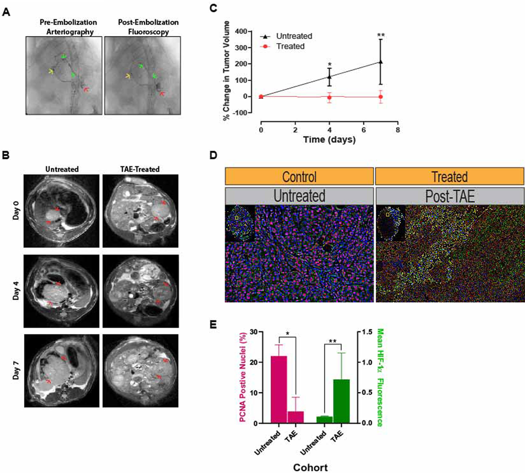
Figure 1. Post-TAE Ischemia Induces Growth Latency In Vivo.
(A) Representative pre-embolization arteriogram demonstrates positioning of the microcatheter in the left hepatic artery (→) with contrast extending to (→) and staining the tumor (→). Post-embolization fluoroscopy demonstrates stasis of contrast within the left hepatic branch artery feeding the tumor as well as persistent contrast staining the tumor. (B) Representative serial T2-weighted MR images of untreated, control HCCs and treated HCCs prior to and following TAE. (C) Growth curves derived from MR-based measurements of tumor volume in untreated HCCs and treated HCCs prior to and following TAE demonstrated a significant reduction in growth for TAE-treated tumors (p=0.011 at posttreatment day 4; p=0.029 at posttreatment day 7)). Tumor volumes are normalized to the first measurement and reported as the average percent change in volume ± standard deviation. (D) Representative immunofluorescence images of untreated control and TAE treated (post-treatment day 7) tumors stained for PCNA (pink) and HIF-1α (green). (E) Bar graph demonstrating a significant reduction in the percentage of DAPI+ nuclei that are also PCNA+ in treated tumors relative to untreated tumors as well as an increase in mean HIF-1α fluorescence intensity per DAPI+ stained nucleus (PCNA: 3.9 ± 4.6% vs 22.0 ± 3.7%, p=.006; HIF-1α: .75 ± .39 vs .11 ± .01, p=.048).

