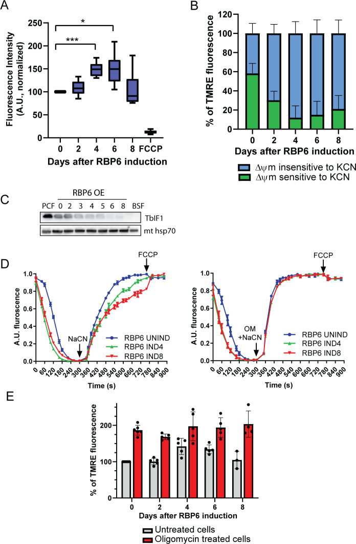
Fig 7. Mitochondrial membrane potential (Δψm) is increased during RBP6OE.
(A) The Δψm of RBP6OE cells post induction was measured by flow cytometry using TMRE. A protonophore FCCP serves as a control for membrane depolarization (mean ± SD, n = 6–10) *P < 0.05, ***P < 0.001. (B) The proportion of Δψm that is generated by complex IV was established by treating the cells with KCN (0.5 mM) in the presence of TMRE for 30 minutes before the analysis. The graph shows a proportion of KCN-sensitive Δψm to the total Δψm measured in each individual sample (mean ± SD, n = 5). (C) Western blot analysis of FoF1-ATPase inhibitory factor TbIF1 during RBP6OE. Mitochondrial (mt) hsp70 serves as a loading control. (D) The in situ dissipation of the Δψm in response to chemical inhibition of complex IV by 1 mM NaCN was measured using safranine O dye in RBP6OE uninduced (UNIND) cells and cells induced for 4 and 6 days. The reaction was initiated with digitonin; OM, oligomycin (2.5 μg/mL), and FCCP (5 μM) were added when indicated (mean ± SD, n = 3). (E) The Δψm of RBP6OE cells that were treated (red columns) or not with oligomycin (2.5 μg/mL). Individual values shown as dots (mean ± SD, n = 3–6). Underlying data plotted in panels A, B, D, and E are provided in S1 Data. BSF, bloodstream cell; FCCP, carbonyl cyanide-4-phenylhydrazone; KCN, potassium cyanide; PCF, procyclic cell; RBP6, RNA binding protein 6; TbIF1, T. brucei inhibitory peptide 1; TMRE, tetramethyl rhodamine ethyl ester.

