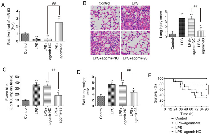
Figure 2.
miR-93 attenuates lung injury in mice with LPS-induced ALI. Mice were injected intravenously with agomir-93 and control agomir (8 mg/kg) 24 h prior to the LPS (2 mg/kg) challenge. Following the LPS administration for 24 h, the mice were sacrificed and the lung tissues were collected for analysis. (A) miR-93 expression was assessed in lung tissues in the model of ALI induced by LPS (n=6 in each group). (B) Staining images of lung tissues following hematoxylin and eosin staining and the pathological changes in lung tissues were evaluated semi-quantitatively based on a histological examination (n=6 in each group). Original magnification, ×100. (C) Lung permeability was assessed using the Evans blue dye extravasation method (n=6 in each group). (D) The lung W/D ratio was determined to evaluate pulmonary edema at 24 h after the LPS challenge (n=6 in each group). Data are the means ± SD (n=3) of one representative experiment, *P<0.05, **P<0.01 vs. the control group. ##P<0.01 vs. the LPS + agomir-NC group. (E) Survival rates were determined using the Kaplan-Meier method. **P<0.01 vs. the LPS alone group. LPS, lipopolysaccharide; ALI, acute lung injury; W/D, wet/dry ratio.

