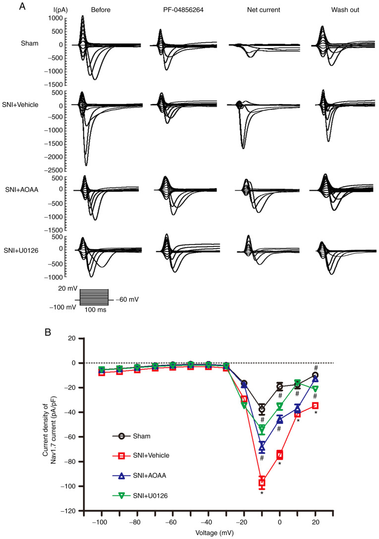
Figure 6.
Changes in the current density carried by Nav1.7 following administration of AOAA or U0126 in DRG neurons of SNI rats. (A) Representative traces showing the sodium current in DRG neurons stimulated using a step protocol from -100 to 20 mV in 10 mV increments with a pulse duration of 100 msec. Before: Traces before cells were perfused with PF-04856264. PF-04856264: Traces after incubation with PF-04856264 for 60 sec. Net current: Traces obtained by subtracting the traces of PF-04856264 from the traces of Before. Wash out: Traces after the extracellular fluid was washed for 4 min. (B) Current density-voltage curves of DRG neurons from each group. *P<0.05 vs. Sham group. #P<0.05 vs. SNI+Vehicle group. n=7 per group. DRG, dorsal root ganglion; SNI, spared nerve injury; AOAA, O-(carboxymethyl) hydroxylamine hemihydrochloride; Nav1.7, voltage-gated sodium channel 1.7.

