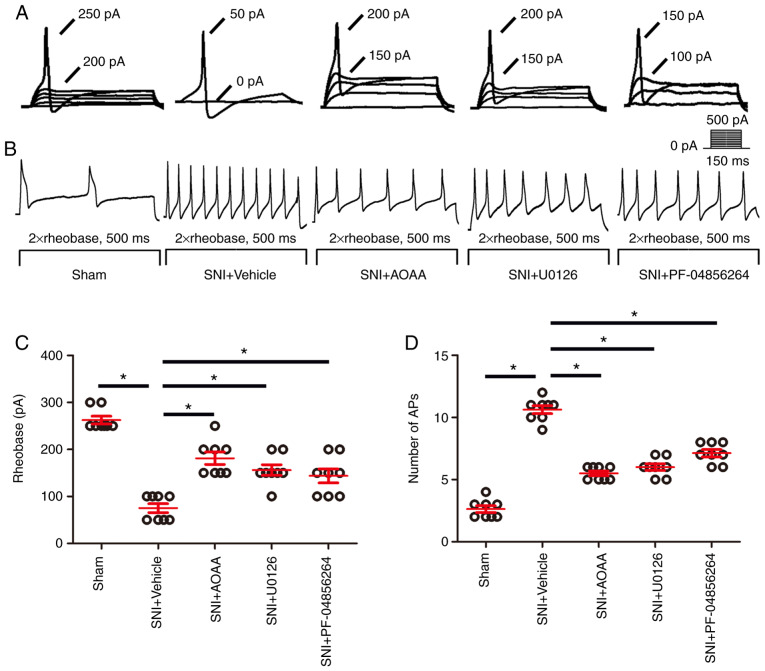
Figure 7.
AOAA, U0126 and PF-04856264 reduce the increase in excitability of DRGs in SNI rats. (A) Representative traces showing the rheobases of action potentials evoked by current injections to DRG neurons of rats in each group. (B) Typical traces showing the action potentials elicited by twice the strength of the rheobase for 500 msec in DRG neurons of rats in each group. (C) Scatter plot showing the statistical comparison of the rheobase of action potentials in each group. (D) Scatter plot showing the statistical comparison of the number of action potentials elicited by twice the strength of the rheobase for 500 msec in each group. *P<0.05. n=8 for each group. DRG, dorsal root ganglion; SNI, spared nerve injury; AOAA, O-(carboxymethyl)hydroxylamine hemihydrochloride. DRG, dorsal root ganglion; SNI, spared nerve injury; AOAA, O-(carboxymethyl) hydroxylamine hemihydrochloride.

