Figure 6.
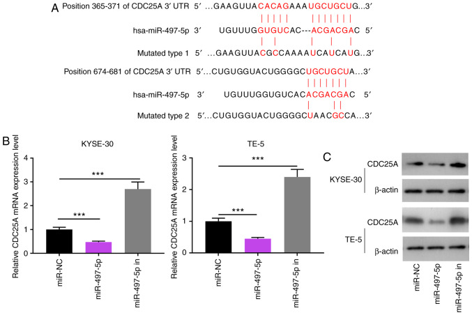
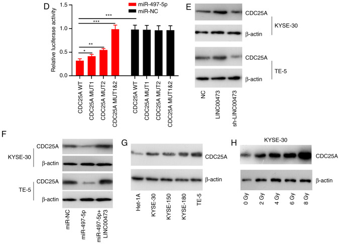
Interaction between miR-497-5p and CDC25A. Binding sites between miR-497-5p and CDC25A were predicted using bioinformatics analysis. (A) Reverse transcription-quantitative PCR was used to quantify the expression of CDC25A miRNA in KYSE-30 and TE-5 cells transfected with miR-NC or miR-497-5p. (B) RT-qPCR analysis of CDC25A expression in KYSE-30 and TE-5 cells transfected with miR-497 mimics or miR-497 inhibitors. (C) Western blot analysis was performed to detect CDC25A expression in KYSE-30 and TE-5 cells transfected with miR-497 mimics or miR-497 inhibitors. (D) The luciferase activity of KYSE-30 and TE-5 cells co-transfected with miR-497-5p and pGL3-miR-497-5p-WT or pGL3-miR-497-5p-MUT. (E) Western blot analysis was performed to detect CDC25A expression in KYSE-30 and TE-5 cells transfected with pcDNA-LINC00473 or sh-LINC00473. (F) Western blot analysis was performed to detected CDC25A expression in KYSE-30 and TE-5 cells transfected with miR-497-5p alone or miR-975-5p+pcDNA-LINC00473. (G) Western blot analysis was performed to detect CDC25A expression in four esophageal squamous cell cancer cell lines (KYSE-30, KYSE-150, KYSE-180 and TE-5) and a normal esophageal epithelial cell line (Het-1A). (H) Western blot analysis was conducted to detect CDC25A expression in KYSE-30 cells under different doses of irradiation. *P<0.05, **P<0.01 and ***P<0.001. miR, microRNA; CDC25A, cell division cycle 25A; NC, negative control; WT, wild-type; MUT, mutant.


