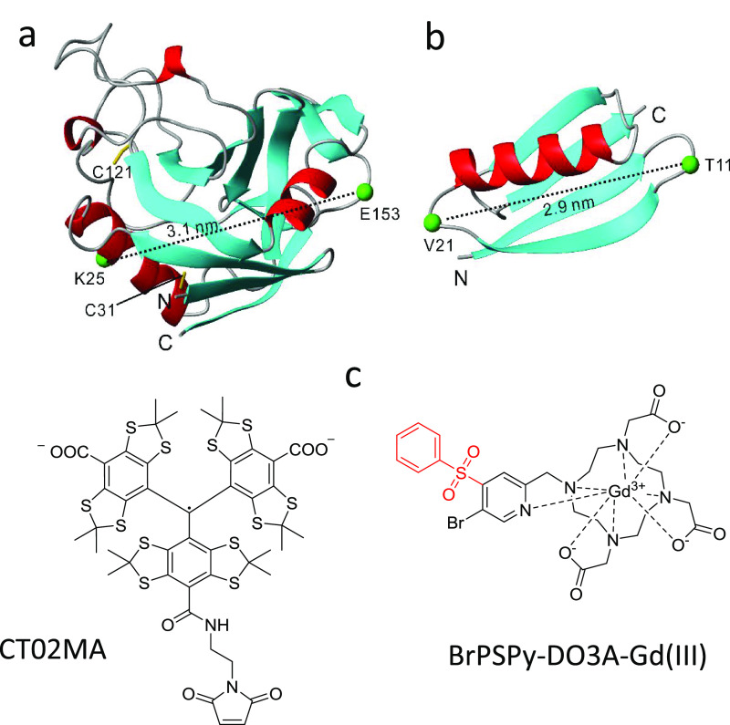
Figure 1.
Structural representation of (a) PpiB and (b) GB1, of which the cysteine mutation at K25 and E153 in PpiB (PDB code: 2NUL(33)) and T11 and V21 in GB1 (PDB code: 2QMT(36)) are shown; the two corresponding Cα atoms, connected by a dashed line, are denoted. The native cysteine residues C31 and C121 in PpiB are relatively buried and were not modified with trityl tags when labeling takes place at 277 K. (c) Molecular structure of the CT02MA and BrPSPy-DO3A-Gd(III) spin labels used in this work.

