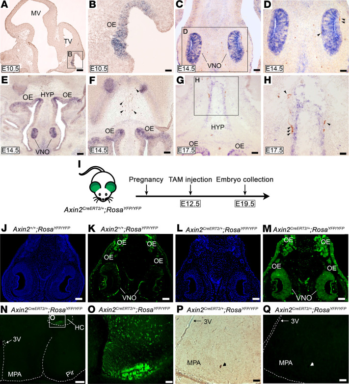
Figure 2. Lgr4/Wnt/β-catenin complex is expressed in key regions for GnRH neuronal development and migration.
(A and B) Low- and high-magnification images of in situ hybridization analysis revealing Lgr4 mRNA localization in the OE at E10.5. (C and D) At E14.5, the signal is strongly visible in the VNO and immunohistochemistry reveals GnRH neurons exiting the VNO. (E and F) Low- and high-magnification images showing Lgr4 mRNA preferentially localized in the VNO, OE, and nuclei of the forebrain at E14.5. (G and H) Low- and high- magnification images showing Lgr4 expression at E17.5 in the OE and in an area of the HYP where GnRH neurons (brown) are migrating alongside. (I) Schematic showing the mouse model employed for lineage tracing. (J and K) Axin2+/+ RosaYFP/YFP control mice reveal a faint and not specific staining for Axin2-GFP. (L and M) In Axin2CreERT2/+ RosaYFP/YFP embryos, Axin2-GFP-positive cells are expressed in the VNO and OE, indicating the presence of Wnt/β-catenin responsive cells. (N and O) Low- and high-magnification images of same specimen showing GFP-positive cells located exclusively in the HC, whereas the MPA is negative. (P) Immunohistochemical detection of GnRH neurons followed by (Q) immunofluorescence for GFP in Axin2CreERT2/+ RosaYFP/YFP embryos. A representative GnRH neuron is GFP negative. Scale bars: 25 μm (A–H), 100 μm (J–M), 250 μm (N), 50 μm (O–Q). Representative images of experiments were performed at least 3 independent times. Arrowheads point to GnRH neurons. 3V, third ventricle; E, embryonic day; HC, hippocampus; HYP, hypothalamus; MPA, medial preoptic area; MV, mesencephalic vesicle; OE, olfactory epithelium; Pir, piriform cortex; TV, telencephalic vesicle; VNO, vomeronasal organ. (A and B) Sagittal sections; (C–Q) coronal sections.

