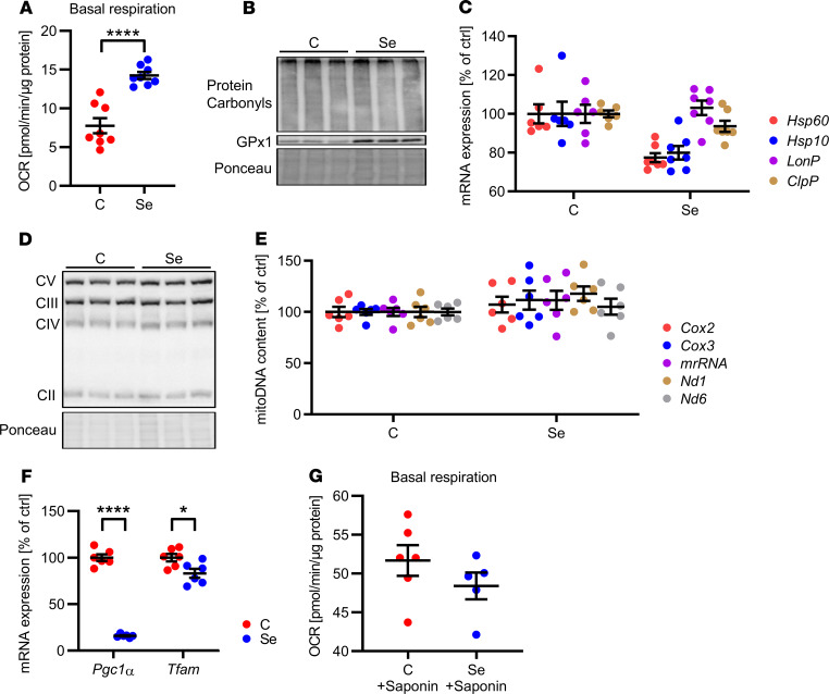
Figure 3. Selenite effect on mitochondrial function in white adipocytes.
(A) Basal oxygen consumption rate (OCR) of 3T3-L1 preadipocytes after selenite treatment using the Seahorse Bioflux Analyzer XF96e (n = 8). (B) Protein carbonylation in 3T3-L1 preadipocytes after selenite treatment. (C) mRNA expression of members of the mitochondrial unfolded protein response in 3T3-L1 preadipocytes after selenite treatment (n = 6). (D) Protein expression of respiratory chain complexes in 3T3-L1 preadipocytes after selenite treatment. (E) Mitochondrial DNA content of 3T3-L1 preadipocytes after selenite treatment (n = 6). (F) mRNA expression of Pgc1a and Tfam in 3T3-L1 preadipocytes after selenite treatment (n = 6). (G) Basal oxygen consumption rate of 3T3-L1 preadipocytes after selenite treatment and permeabilization with saponin using the Seahorse Bioflux Analyzer XF96e (n = 7). *P < 0.05, and ****P < 0.0001 after 2-tailed Student’s t test. All data are presented as mean ± SEM.

