. 2020 Jun 4;9:e58130. doi: 10.7554/eLife.58130

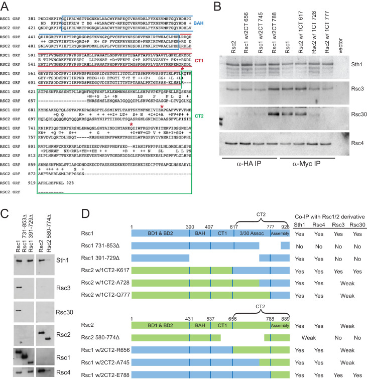
Figure 1. Rsc3/30 module has higher avidity for the RSC1 complex.

(A) Purified RSC1-TAP and RSC2-TAP complexes (2 µg) analyzed on 7.5% SDS-PAGE gel stained with Coomassie dye. (B) RSC1 and RSC2 complex compositions, with decreased opacity displaying the reduced association of the Rsc3/30 module in the RSC2 complex. (C) Domain structure and swaps of Rsc1 and Rsc2. (D) CT2 domain swaps complement for viability. TRP1-marked plasmids bearing RSC1 (p609), RSC2 (p604), RSC1 w/2CT2 (p3097), RSC2 w/1CT2 (p3098), or vector (pRS314) were transformed into rsc1rsc2∆ [RSC1.URA3] (YBC800), and spotted as 10x serial dilutions to SC-TRP or to SC-TRP+5FOA to force the loss of the RSC1.URA3 plasmid. ’ w/’ indicates ‘with’. One of four biological replicates shown. (E) The Rsc3/30 module associates more strongly with the CT2 region of Rsc1. Immunoprecipitations of Rsc1, Rsc2 w/1CT2, Rsc2, Rsc1 w/2CT2 from whole cell extracts. Blots were probed with anti-Sth1, then stripped and reprobed for anti-Rsc3 and anti-Rsc4. One of three technical replicates shown. Figure 1—figure supplement 1. The Rsc3/30 module associates with the RSC1 complex at high stringency. Figure 1—figure supplement 2. Additional swaps and truncations define the region of Rsc3/30 association.

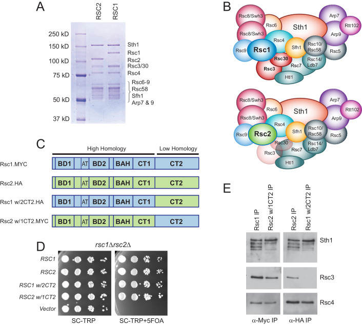
Figure 1.

Figure 1—figure supplement 1. The Rsc3/30 module associates with the RSC1 complex at high stringency.

Figure 1—figure supplement 1.

Yeast cell lysates containing RSC-TAP complexes were bound to IgG resin, the beads rinsed with lysis buffer before washing with buffer of the indicated salt concentration and duration. RSC complexes were analyzed on 6% acrylamide SDS-PAGE gels stained with silver. The Rsc3 and Rsc30 proteins run closely on denaturing gels and the doublet will often appear as a single band, marked by the asterisk (*). The marker lanes (M) are each from the adjacent gel, but were moved for depiction. Stringency testing conducted once.
Figure 1—figure supplement 2. Additional swaps and truncations define the region of Rsc3/30 association.
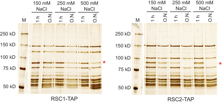
Figure 1—figure supplement 2.

(A) Alignment of C-terminus of Rsc1 and Rsc2, showing conservation and position of swaps (red asterisks). (B) Co-IPs of Rsc3 and Rsc30 with additional Rsc1 and Rsc2 CT swaps. Whole cell extracts prepared from rsc1rsc2∆ covered by TRP1-marked plasmid containing RSC1, RSC2, or CT swap were immunoprecipitated using anti-Myc (for CT2 of Rsc1), or anti-HA (for CT2 of Rsc2) bound to Dynabeads. Western blots were probed with antibodies against Sth1, Rsc3, Rsc30, and Rsc4. One of three biological replicates shown. (C) Co-IPs of RSC members with Rsc1 or Rsc2 containing deletions within the CT2 region. Whole cell extracts prepared from rsc1rsc2∆ covered by TRP1-marked plasmid containing deletions were immunoprecipitated using anti-Myc (for CT2 of Rsc1), or anti-HA (for CT2 of Rsc2) bound Dynabeads. Western blots were probed with antibodies against Sth1, Rsc3, Rsc30, Rsc2, Rsc4, and Myc. One of three technical replicates shown. (D) Summary of Rsc1 and Rsc2 Co-IPs from deletions and swaps.