. 2020 Jun 4;9:e58130. doi: 10.7554/eLife.58130

Figure 3. Mutations in the H3 αN helix are lethal in combination with rsc1∆, but not rsc2∆, and they reduce RSC remodeling of the 5S nucleosome.

(A) Histone H3 αN helix mutations that are lethal with rsc1∆. TRP1-marked plasmids containing WT H4, and H3 mutations within the αN helix were transformed into h3-h4∆ [H3-H4.URA3] (YBC1939), rsc1h3-h4∆ [H3-H4.URA3] (YBC2090) or rsc2h3-h4∆ [H3-H4.URA3] (YBC3040), and spotted to SC-TRP or SC-TRP+5FOA to force the loss of the WT histone plasmid. Mutations that were lethal on their own without mutated RSC are shaded in grey. Mutations that were lethal in rsc1∆ but not rsc2∆ are shaded in purple and underlined. Transformants were grown at 30°C for 2 days. Shown is one of two biological replicates. (B) Location of the synthetic lethal rsc1∆ H3 αN helix mutations are depicted in purple on the nucleosome, PDB code 1ID3. (C) The RSC1 CT2 region complements the synthetic lethal rsc1∆ H3 αN helix mutations. rsc1rsc2h3-h4∆ [RSC1.URA3] with [H3.WT, R40A, or G44A-H4.WT. LYS2] (YBC3466, YBC3444, YBC3433) transformed with TRP1-marked plasmids bearing RSC1 (p609), RSC2 (p604), RSC1 w/2CT2 (p3097), RSC2 w/1CT2 (p3098), or vector (pRS314) and spotted as 10x serial dilution to SC-TRP-LYS, or SC-TRP-LYS+5FOA. Shown is one of four biological replicates. (D) High-copy RSC3 or RSC30 will partially suppress the Tsˉ phenotype of rsc1H3V46A. Strain rsc1h3-h4∆ [H3.V46A-H4.WT.TRP] (YBC3586) transformed with URA3-marked high copy (2μ) plasmids containing RSC1 (p705), RSC3 (p1310), RSC30 (p916), HMO1 (p3390), or vector (pRS426), and spotted as 10x serial dilutions at 30°C or 37°C. Shown is one of two biological replicates. (E) Comparative sliding and ejection of 174 bp sea urchin 5S NPS H3 R40A yeast mononucleosomes (20 nM) by RSC1 and RSC2 complexes (30 nM). The nucleosomal Start (green), Slid (blue), and free DNA (grey) bands were quantified and reported as a percent of the total signal.

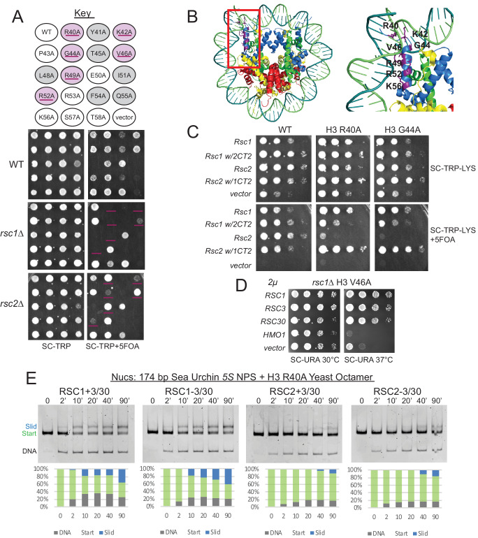
Figure 3—source data 1. RSC sliding 5S R40A yNuc.

Figure 3.

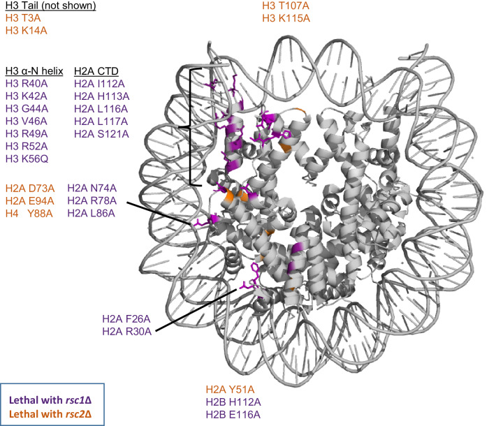
Figure 3—figure supplement 1. Synthetic lethal mutations in each of the four histones H2A, H2B, H3, and H4 when combined with rsc1∆ or rsc2∆ mutations.

Figure 3—figure supplement 1.

Mutations that are specifically lethal in combination with rsc1∆ are colored purple, mutations that are specifically lethal with rsc2∆ are colored orange. PDB code 1ID3.
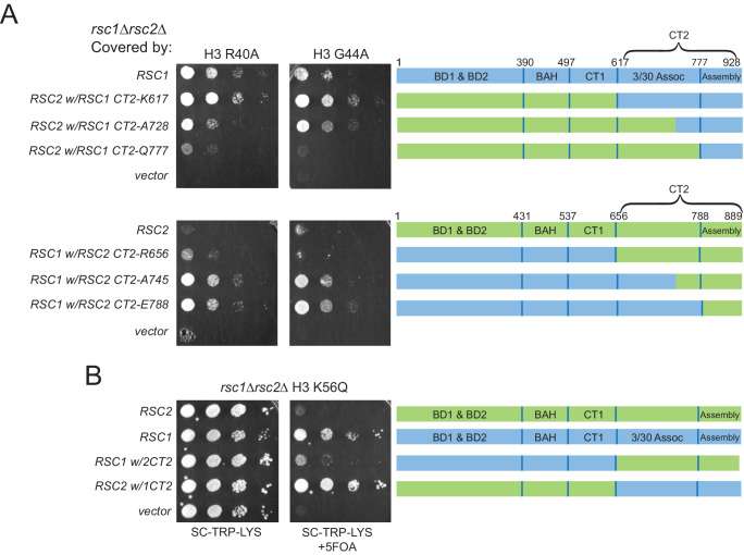
Figure 3—figure supplement 2. Complementation by CT2 domain swaps when combined with H3 αN helix mutations.

Figure 3—figure supplement 2.

(A) The RSC1 CT2 region complements the synthetic lethal rsc1∆ H3 αN helix mutations. Strain rsc1rsc2h3-h4∆ [RSC1.URA3] [H3. R40A, or G44A-H4.WT. LYS2] (YBC3444, YBC3433) transformed with TRP1-marked plasmids bearing RSC1 (p609), RSC2 (p604), C-Terminal swaps at positions noted (p3098, p3165, p3167, p3097, p3166, p3164) or vector (pRS314) and spotted as 10x serial dilution to SC-TRP-LYS, or SC-TRP-LYS+5FOA. One of four biological replicates shown. (B) Synthetic lethality of rsc1∆ H3 K56Q is complemented by the RSC1 CT2 region. Strain rsc1∆ rsc2∆ h3-h4∆ [RSC1.URA3] [H3.K56Q-H4.WT. LYS2] (YBC3432) transformed with TRP1-marked plasmids bearing RSC1 (p609), RSC2 (p604), RSC1 w/2CT2 (p3097), RSC2 w/1CT2 (p3098), or vector (pRS314) and spotted as 10x serial dilution to SC-TRP-LYS, or SC-TRP-LYS+5FOA. One of four biological replicates shown.
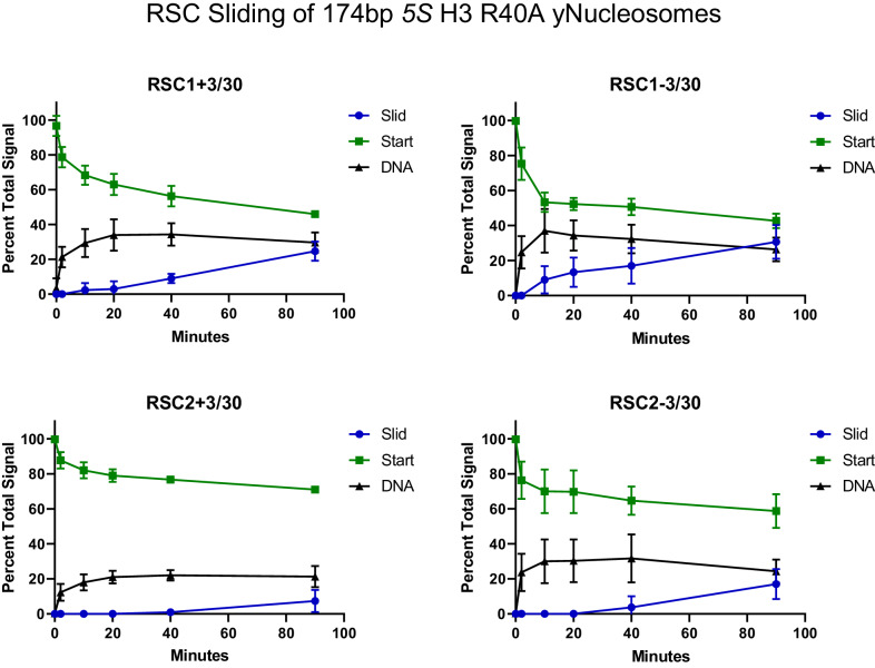
Figure 3—figure supplement 3. Quantification of RSC sliding 174 bp 5S H3 R40A yeast mononucleosomes as conducted in Figure 3E.

Figure 3—figure supplement 3.

Mean and standard deviation for three replicates.
Figure 3—figure supplement 3—source data 1. Quantification of RSC sliding 5S R40A yNucs.
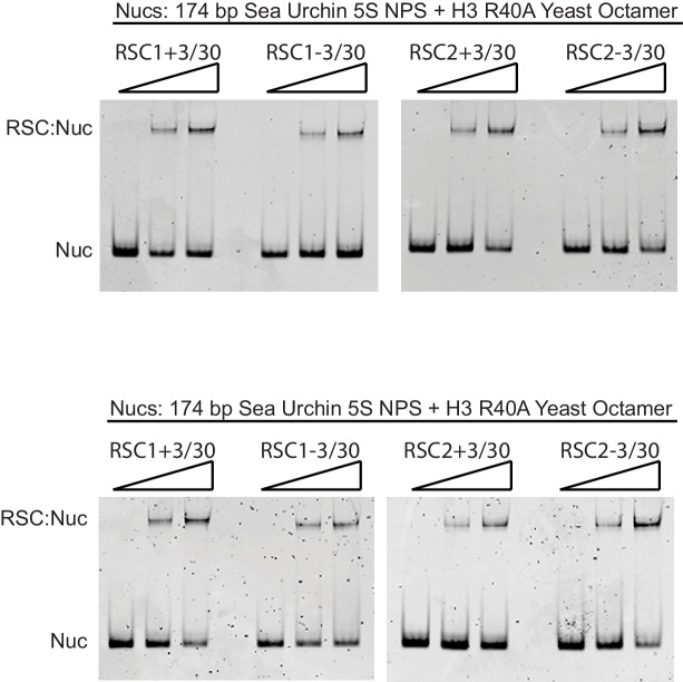
Figure 3—figure supplement 4. RSC1 and RSC2 complexes bind similarly to 174 bp sea urchin 5S yeast mononucleosomes with the H3 R40A mutation.

Figure 3—figure supplement 4.

Nucleosomes (20 nM) were incubated with 0, 30, or 60 nM RSC for 20 min at 30°C, run on a 3.8% native polyacrylamide gel, and stained with ethidium bromide. Two technical replicates shown.
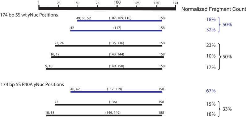
Figure 3—figure supplement 5. Mapping the positions of the wt and H3 R40A 174 bp sea urchin 5S yeast mononucleosomes.

Figure 3—figure supplement 5.

Nucleosome mapping by ExoIII and S1 treatment followed by paired-end next-generation sequencing. Experiment conducted once. Partially wrapped nucleosome fragments (≤120 bp) are depicted in blue. More fully wrapped nucleosome fragments (≥128 bp) are in black.
Figure 3—figure supplement 5—source data 1. 174 bp 5S nucleosome mapping.