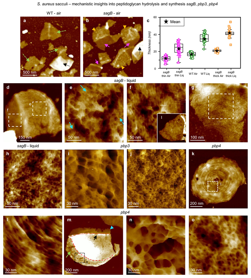
Extended Data Figure 3. Mechanistic insight into peptidoglycan hydrolysis and synthesis.
a, Sacculi of S. aureus WT imaged in air, with different fragments containing single layers of PG (green dashed arrows) and double layers (black dotted arrow), DS = 75 nm. b, Sacculi from S. aureus sagB mutant in an SH1000 background imaged in air. The fragments have two distinct regions with different thicknesses (pink and orange arrow heads). These are distinct from the double layer fragments (black dotted arrow), DS = 75 nm. c, Graph comparing the thickness of the cell wall in air and liquid between the sagB mutant and the wild type. The box-plot consists of boxes from 25-75 percentile, middle line represents the median, black star represents the mean value, error bars are maximum/minimum values excluding any outliers (using 1.5 inter-quartile range). The two different regions of the sagB cell wall are separately binned and labelled ‘sagB thin’ and ‘sagB thick’. Measurements were taken using the same approach as the values for graph Fig 2k in the main text and ED4. Number of sacculi measured n = 25 for each bin. Mean values ± s.d. are: ‘sagB thin air’ = 12 ± 3 nm, ‘sagB thin liq’ = 24 ± 7 nm, ‘sagB thick air’ = 21 ± 1.5 nm, ‘sagB thick liq’ = 42 ± 5.7 nm, ‘WT air’ and ‘WT liq’ corresponded to the same values as Fig 2k (left data, PG+WTA). All measurements in liquid were performed in buffer 300 mM KCl + 10 mM Tris, pH = 8. d, S. aureus sagB mutant sacculus in liquid with its external surface facing upwards. This sacculus is clearly divided into two sections, the region on the left is thinner than the region on the right. This irregularity in height was visualised in several sacculi from cells with this mutation. DS = 64 nm. e, Higher resolution image from the thicker section in d corresponding to the finer mature CW architecture. This mesh is still in an early transition from rings, with some features reminiscent of rings visualised following the same orientation (blue arrows). These sorts of structures are uncommon on WT sacculi at the same stage in the cell cycle (exponential phase OD600 = 0.5-0.7). However, in the sagB mutant it was more common to find this early transition from rings to mesh. DS = 11 nm. f, Higher resolution image from the thinner section in d showing the finer nascent CW architecture (i.e. rings). Inset shows a lower resolution image of the ring architecture in a different sacculus. The features from this image are not significantly different from WT nascent PG architecture, such as Fig 2d in the main text. However, almost every sacculus containing nascent architecture in the sagB mutant had this part of the cell wall being thinner than the rest of the sacculus, something not seen in the WT. DS = 9 nm, inset DS = 29 nm. g, Sacculus fragment with the inner CW architecture facing upwards, DS = 14 nm. h, Higher resolution image from g showing the fine inner structure of the sagB mutant, with no clear difference from the very tight and disordered structure seen in WT sacculi (see Fig 2f from the main text for example). DS = 4 nm. i, Sacculi from S. aureus pbp3 mutant cells in an SH1000 background. Here we show the fine external structure, with no apparent difference between this and the S. aureus WT mature external structure. It is important to highlight that PBP3 is not an essential peptidoglycan synthesis enzyme, and the cells can survive without it and still maintain their spheroid shape. DS = 55 nm. j, The fine structure of an S. aureus pbp3 mutant sacculus with the internal peptidoglycan surface exposed, which is similar to that seen in wild type S. aureus. DS = 4 nm. k, Sacculus from S. aureus pbp4 mutant cells in an SH1000 background showing nascent peptidoglycan structure, with the concentric rings facing upwards. It is important to highlight that PBP4 is not an essential peptidoglycan synthesis enzyme either, and the cells can survive without it and still maintain their spheroid shape. DS = 28 nm. l, Higher resolution image taken within k showing the fine structure of the nascent peptidoglycan surface, the concentric rings. There is no significant difference compared to the S. aureus WT nascent structure, except the fraction of sacculi with rings in a random area is higher in this sample than WT. DS = 8 nm. m, Sacculus showing a small area of the mature peptidoglycan structure (green long arrow), with its external surface facing upwards, the rest of the sacculus is covered by concentric rings already in the late phase of the transition to mesh (black short arrow pointing to the centre of the concentric rings, boundary with the mature structure, red dashed line) and the newest generation fragment with rings (blue arrow head, boundary with oldest rings marked with red dotted line). It is important to highlight this is the first time two perpendicular planes of rings have been visualized at exponential phase in our experience. OD600= 0.6. DS = 79 nm. n, Higher resolution image from m showing the fine structure of the mature external CW of an S. aureus pbp4 mutant, with no apparent difference between this and the S. aureus WT mature external structure (see ED1 and Fig 2b from the main text). DS = 44 nm. o, Sacculus showing the fine internal structure of S. aureus pbp4 mutant cell wall, with no apparent difference compared to the S. aureus WT internal peptidoglycan architecture (see Fig 2f from the main text). DS = 10 nm. These data show that removal of non-essential PBPs makes only relatively minor differences to the wall architecture.

