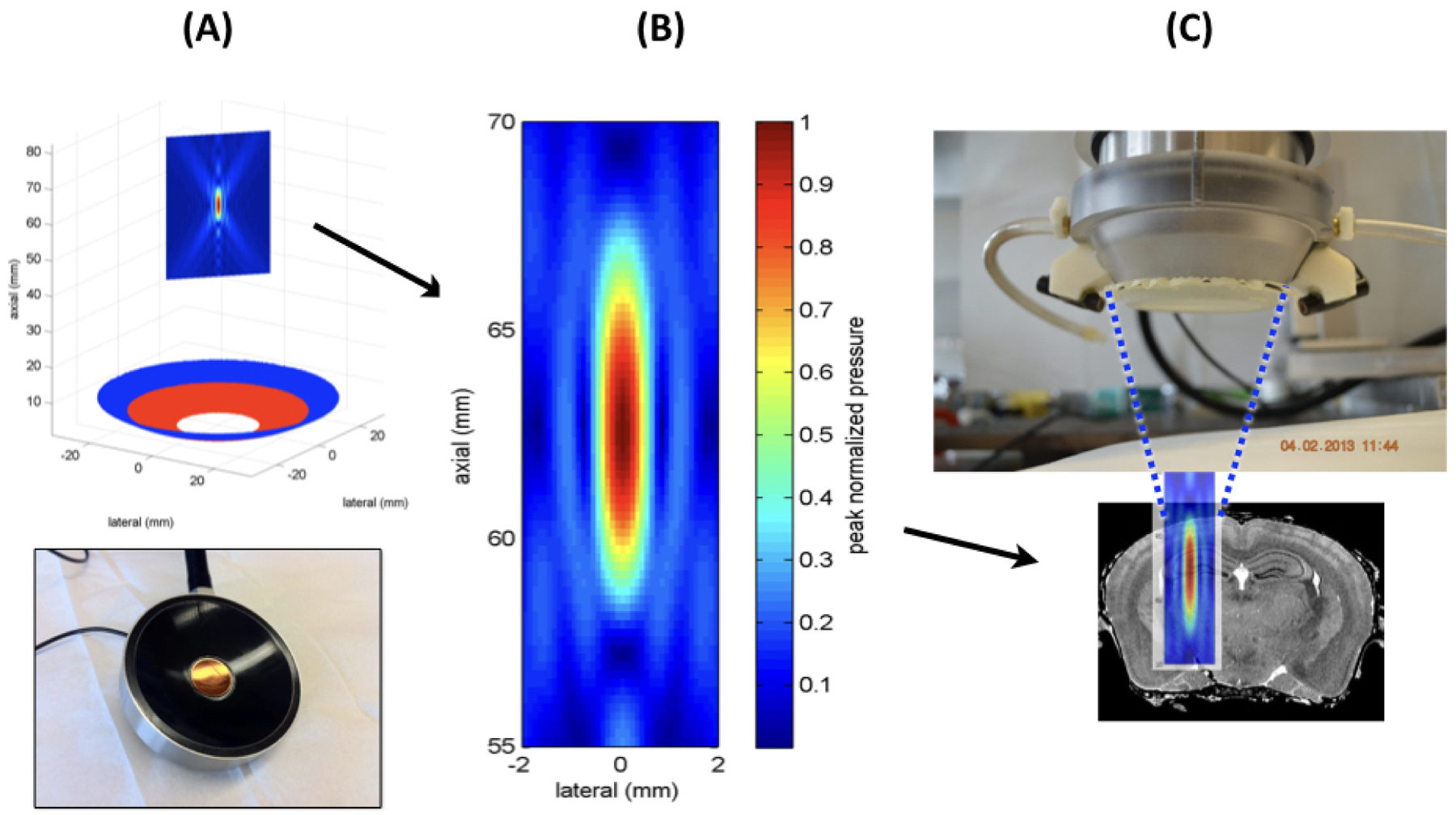
Figure 1. Montage of our experimental tFUS device.

(A) This figure shows the transducer portion of the entire tFUS device below a mathematical representation of the two-annular transducer (red and blue circles) beneath a planar representation of the focal volume of ultrasound. (B) This figure shows a closeup of a mathematical representation of the maximum pressure field generated within the focal region of our ultrasound source as if it were measured in water. (C) This figure shows the working end of the tFUS device (top) with water-filled cone and closing membrane over ultrasound field of (B) itself superimposed upon a coronal slice of mouse brain, to scale.
