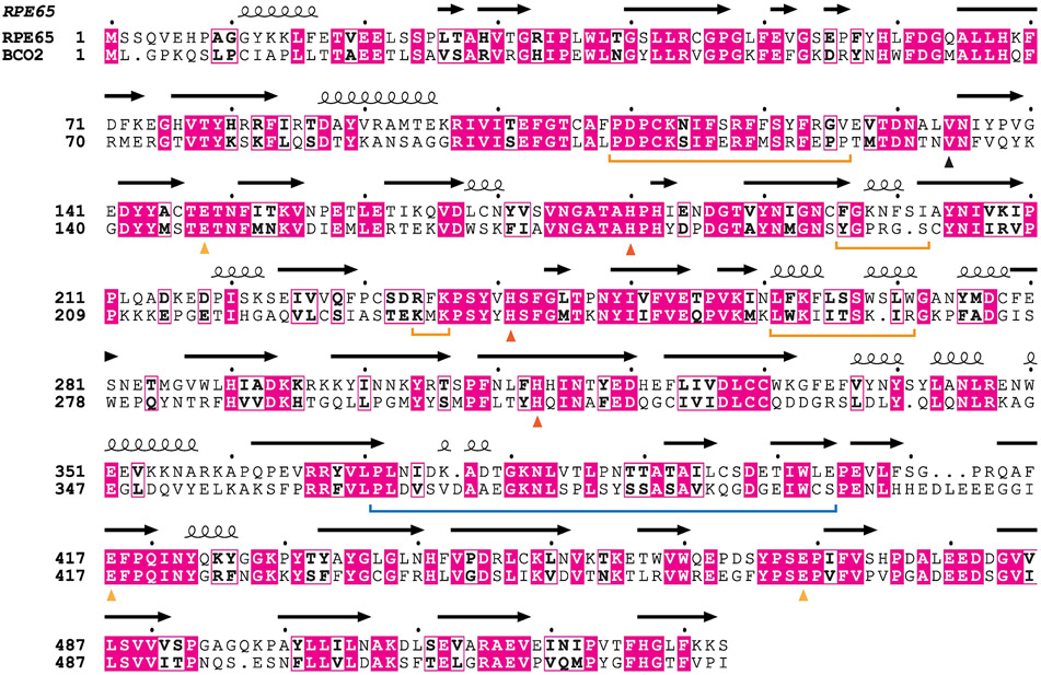
Figure 10. Sequence alignment between bovine RPE65 and mouse BCO2.
Secondary structure for RPE65 determined by crystallography is shown above the alignment. Residues thought to mediate membrane binding of these proteins are marked by horizontal orange brackets. The region mediating dimer formation in RPE65 is marked by a horizontal blue bracket. The conserved metal-binding His and Glu residues are marked by red and orange triangles, respectively, while the occluding Val residue is marked by a black triangle.

