. 2020 Mar 20;38(3):323–337. doi: 10.5534/wjmh.200012

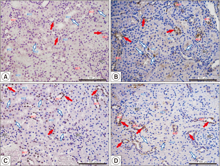
Fig. 4. Representative microphotography showing terminal deoxynucleotidyl transferase dUTP nick end labeling reaction (A, C) and proliferating cell nuclear antigen-positive cells (B, D) in control (A, B) and finasteride-treated (C, D) rats. Red filled arrows indicate positive signaling in nuclei of proximal convoluted tubule (PCT); blue empty arrows indicate positive signaling in nuclei of the distal convoluted tubule (DCT). Slides were rinsed in phosphate buffer saline and labeled with streptavidin conjugated with horseradish peroxidase. To visualize the effect of the reaction (places of DNA split), 3,3′-diaminobenzidine was added. Positive staining was defined microscopically (Leica DM5000B, Germany) through visual identification of brown pigmentation of the cell nucleus. Scale bar from objective magnification ×40 (A, B; C, D) is 100 mm. RC: renal corpuscle. Data from Baig et al (Int J Environ Res Public Health 2019;16:E1726) [18].

Fig. 4