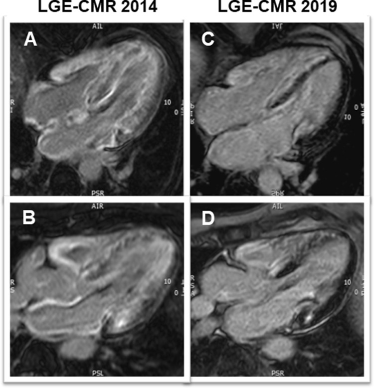
Fig. 3.
Late gadolinium enhancement (LGE) CMR images in four- (a, c) and three-chamber (b, d) views from first CMR (2014; a, b) and at present evaluation (2019; c, d). At first CMR (a, b) extensive, circumferential (non-ischemic pattern) LGE in both ventricles a well as in the walls of both atria—a characteristic finding for cardiac amyloidosis—can be seen. At present evaluation (c, d), an obvious decrease in the LGE extent, particularly in the lateral wall of the LV, in the subendocardium of the septum and in the atrial walls is noted

