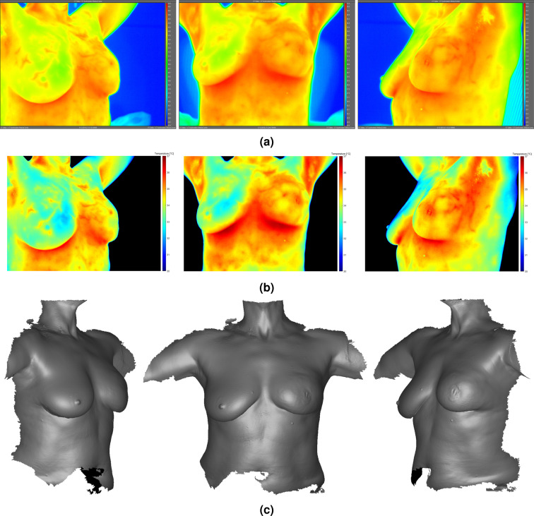
Figure 3.
Infrared images (a,b) and 3D breast surface scans (c) of Subject 03. IR images in (a) were exported directly from FLIR ResearchIR Max; IR images in (b) were processed using MATLAB to modify background color to black. Clear thermovascular asymmetry was observed between breasts due to generalized hyperthermia in subject’s left breast (see Table 2 for summary of temperatures). Physical deformity in subject’s left breast due to malignancy was evident at presentation during research procedures. High-resolution IR images available for download online (see Supplementary Figs. S1–S6).

