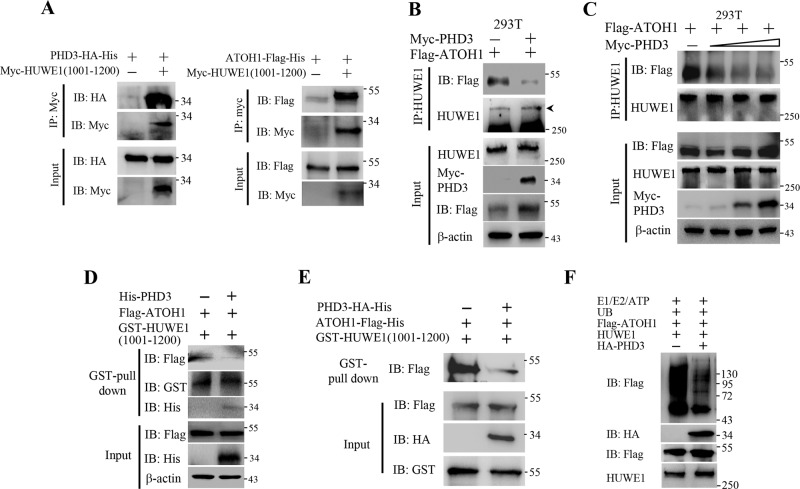
Fig. 4. PHD3 blocked the HUWE1–ATOH1 interaction.
a 293T cells were transfected with myc-HUWE1(1001–1200) vector. After 24 h, cell lysates (200 μg) were prepared and incubated with 1 μg Myc antibody at 4 °C for 3 h, followed by incubation with 40 μl glutathione-Sepharose beads for another 1 h. The beads were washed and 30 μg purified PHD3-HA-His or ATOH1-Flag-His proteins were added and incubated at 4 °C overnight. The beads were washed, boiled in SDS-PAGE loading buffer, and the resolved proteins were subjected to western blot. b 293T cells were transfected with Myc-PHD3 or Myc-PHD3 plus Flag-ATOH1 vector. Twenty-four hours post transfection, the cells were treated with MG132 (10 μM) for 4 h, followed by immunoprecipitation experiment. c 293T cells were transfected with Flag-ATOH1 and different amount of Myc-PHD3 (0, 0.5, 1 or 2 μg) vector. After 24 h, the cells were treated with MG132 (10 μM) for 4 h, followed by immunoprecipitation assay. d The beads that captured GST-HUWE1(1001–1200) were incubated at 4 °C with 293T lysates containing Flag-ATOH1 for 2 h. The beads were washed and incubated with bacteria lysates containing His-PHD3 at 4 °C overnight. e GST-HUWE1(1001-1200) was incubated with glutathione-Sepharose beads at 4 °C for 1 h. The beads were washed and incubated with ATOH1-Flag-His (18 μg) or equal amount of ATOH1-Flag-His plus PHD3-HA-His at 4 °C overnight. The beads were washed, boiled in SDS-PAGE loading buffer, and the resolved proteins were detected by immunoblotting. f Purified PHD3-HA-His inhibited the ubiquitination of ATOH1-Flag-His by HUWE1 in vitro. The experiment was performed as described in “Methods”.

