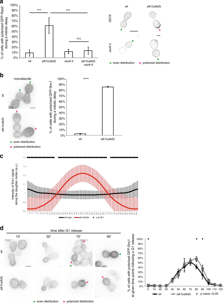
Fig. 5. Haspin promotes a shift from apical- to whole PM-oriented vesicle delivery required for Ras dispersion.
a Cells of the indicated strains were grown at permissive temperature (25 °C) in raffinose-containing medium. After G1-synchronization, strains were released in nocodazole containing medium supplemented with 2% galactose to induce expression of GFP-RAS2. After 2.5 h, cultures were shifted to 37 °C to inactivate exocytosis for further 1.5 h. Samples were then taken and analyzed by fluorescence microscopy. The graph represents the percentage of cells with polarized GFP-Ras2; representative exhibits are shown on the right, green and magenta arrows indicate cells with even or polarized GFP-Ras2 signal, respectively. wt or haspin-lacking cells expressing GFP-SNC1 were arrested in G1 and then released for 2.5 h in nocodazole-containing medium b and c or released in fresh medium without drugs d. Samples were analyzed by fluorescence microscopy. Representative images are shown, and the quantified results are reported in the graphs. Green and magenta arrows indicate cells with even or polarized Snc1, respectively. For graphs a, b, d three experiments were performed counting 100 cells per repeat, error bars represent standard deviation. Cdc24-GFP signal intensity along the PM quantified on 60 nocodazole-arrested cells from three experiments were performed, counting 100 cells for each repeat; error bars represent standard deviation. Scale bars in a, b, d: 5 μm. t-test was applied as a statistical measurement in a–d; n.s.: not significant; *P < 0.05; **P < 0.01; ***P < 0.005; ****P < 0.001.

