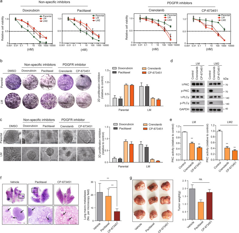
Fig. 3. Preferential anti-metastasis efficiency of PDGFR inhibition.
a Cell viability of parental, LM1, and LM2 cells upon treatment with increasing concentrations of doxorubicin, paclitaxel, crenolanib, or CP-673451. b Colony-formation assays of parental or LM cells treated with DMSO, doxorubicin (50 nM), paclitaxel (10 nM), crenolanib (1 μM), or CP-673451 (1 μM). Cells were cultured for 8 days followed by staining (left) and quantification (right). Scale bars, 100 µm. c 3D-Matrigel on-top assays of parental or LM cells treated with DMSO, doxorubicin (50 nM), paclitaxel (10 nM), crenolanib (1 μM), or CP-673451 (1 μM). Representative images (left) and quantification (right) of 3D-organoid formation after 10 days of culture. Scale bars, 100 µm. d LM1 and LM2 cells were treated with DMSO, crenolanib (1 µM), or CP-673451 (1 μM) for 48 h, and changes in the total PKC, p-PKC, total PLCγ, and p-PLCγ protein levels were analyzed by western blot. GAPDH served as a loading control. e Levels of total PKC kinase activity in LM1 and LM2 cells treated with crenolanib (1 μM) or CP-673451 (1 μM) relative to DMSO-treated cells are shown. f Representative H&E staining (left) and quantification (right) of lung macro-metastases of LM tumor-bearing mice treated with vehicle, paclitaxel (10 mg/kg), or CP-673451 (50 mg/kg). Scale bars, 200 µm. g Photographs of harvested tumors (left) and quantification (right) of primary tumor weight of LM tumor-bearing mice treated with vehicle, paclitaxel (10 mg/kg), or CP-673451 (50 mg/kg).

