
Fig. 2. Sibling preference in young pups is disrupted by lesion of the lateral septum.
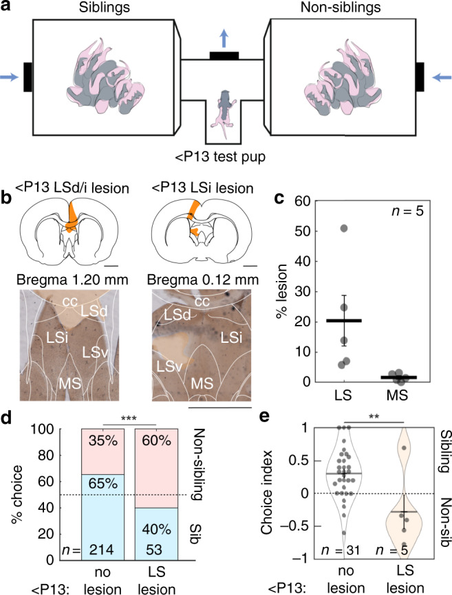
a Apparatus for sibling-preference testing. Test pup was placed in the T-tube with two opposing chambers containing sibling and non-sibling rats. Air was blown toward the center of the T-choice tube. b Examples of two sections from two pups with lateral septal lesions. Left, lesion of dorsal and intermediate lateral septum (LSd/i) with overlying cortex. Right, lesion of intermediate lateral septum (LSi) and cortex (lesion area in orange). Scale bar applies to left and right micrographs. c Sections (100 µm thickness) of the five lesioned brains were analyzed for lesion extent. Average percent lesion of septum was estimated (LS: 20.45 ± 8.36%; MS: 1.53 ± 0.56%). Data represent the mean ± s.e.m. d Sibling preference in intact animals and with lateral septum lesion. Bars represent proportions of categorical choices pooled across animals (<P13 No lesion (as in Fig. 1d): 140 sibling choices, 74 non-sibling choices; <P13 lateral septum lesion: 21 sibling choices, 32 non-sibling choices; p = 8.9e−4, Fisher’s exact test). e Sibling choice behavior by animal, non-lesioned data as in Fig. 1e (<P13 No lesion vs. LS lesion: p = 0.005, t-test). Data represent the mean ± s.e.m. All tests are two-tailed. For detailed statistical information, see Supplementary Table 1. LSd dorsal lateral septum, LSi intermediate lateral septum, LSv ventral lateral septum, cc corpus callosum, MS medial septum. Scale bars: 2 mm. **p < 0.01, ***p < 0.001.