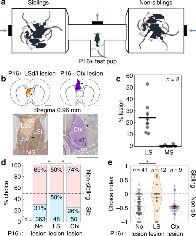
Fig. 3. Non-sibling preference of old pups is disrupted by lesion of the lateral septum.

a Apparatus for sibling-preference testing. Test pup was placed in the T-tube with two opposing chambers containing sibling and non-sibling rats. Air was blown toward the center of the T-choice tube. b Examples of two sections from two pups with lateral septal and cortical lesions (LS: lesion area in orange, Ctx: lesion area in purple). Scale bar applies to left and right micrographs. c Sections (100 µm thickness) of eight lesioned brains were analyzed for lesion extent. Four septal lesioned brains could not be measured due to damage during processing. Average percent lesion of lateral septum was estimated (LS: 24.09 ± 4.82%; MS: 0.46 ± 0.31%). Data represent the mean ± s.e.m. d Comparison of sibling preference in no lesion, lateral septum lesion and cortex lesion pups. Bars represent proportions of categorical choices pooled across animals (P16+ no lesion (as in Fig. 1d): 112 sibling choices, 251 non-sibling choices; P16+ lateral septal lesion: 24 sibling choices, 24 non-sibling choices; P16+ Ctx lesion: 13 sibling choices, 37 non-sibling choices; no lesion vs. LS lesion: p = 0.014; LS lesion vs. Ctx lesion: p = 0.021; no lesion vs. Ctx lesion: p = 0.517; Fisher’s exact test). e Sibling choice behavior by animals. Data represent the mean ± s.e.m. (No lesion vs. LS lesion: p = 0.012, t-test; no lesion vs. Ctx lesion: p = 0.893, t-test; LS lesion vs. Ctx lesion: p = 0.103, t-test; no lesion as in Fig. 1e). All tests are two-tailed. For detailed statistical information, see Supplementary Table 1. LSd dorsal lateral septum, LSi intermediate lateral septum, LSv ventral lateral septum, cc corpus callosum, MS medial septum, Ctx cortex. Scale bars: 2 mm. *p < 0.05.
