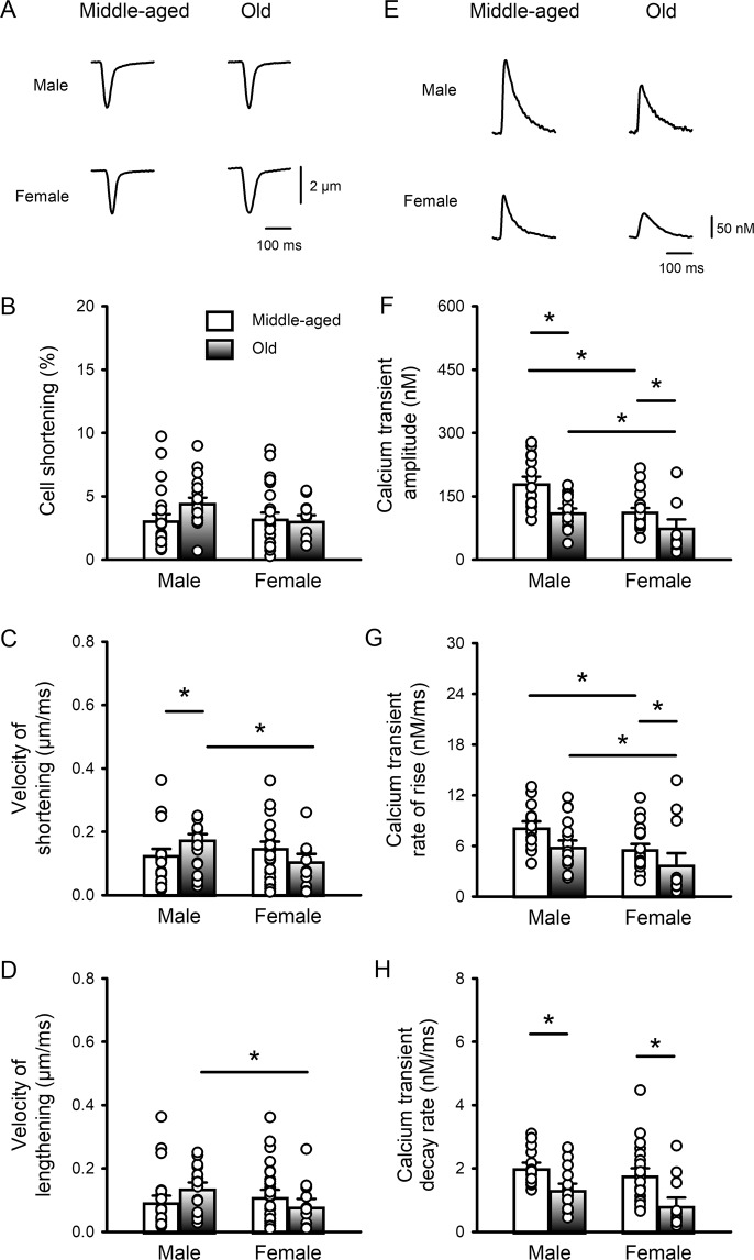
Figure 1.
Peak calcium transients declined and slowed with age in C57BL/6 mice of both sexes, but contractions were largely unaffected. (A) Representative examples of contractions (cell shortening) recorded from field-stimulated (4 Hz) ventricular myocytes isolated from middle-aged (~12 mos) and older (~24 mos) male and female mice. (B) Mean data show that peak contractions were similar in all four groups. (C) The velocity of shortening increased slightly with age in males and was faster in older male cells compared to female cells. (D) The velocity of lengthening was unaffected by age but was lower in older females than in older males. (E) Representative examples of calcium transients recorded from myocytes from middle-aged and older mice of both sexes. (F) Mean data show that peak calcium transients declined with age in both sexes and were smaller in cells from females than males at both ages. (G) The rates of rise of the calcium transient declined with age and this was significant in females. The rates of rise were slower in females than males at all ages. (H) The rates of decay of the calcium transients declined markedly with age in both sexes. Values represent the mean ± SEM values in each case. Data were analyzed by two-way ANOVA with age and sex as main factors (post-hoc test was Holm-Sidak). The * denotes p < 0.05. For calcium transients n = 15, 18, 22 and 12 myocytes from 6 middle-aged male mice, 4 older males, 9 middle-aged females and 3 older females, respectively. For contractions n = 24, 20, 27 and 12 myocytes from 6 middle-aged male mice, 4 older males, 8 middle-aged females and 3 older females, respectively.

