
FIGURE 1.
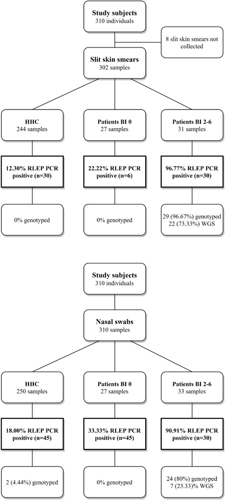
Study design, RLEP positivity and genotyped samples. Flow diagram providing an overview of the subjects recruited for this study. Slit skin smears (SSS) and nasal swabs (NS) collected per group; healthy household contacts (HHC), paucibacillary (PB) or multibacillary (MB) patients with BI 0, and MB patients with a bacteriological index (BI) 2–6. MB patients with BI 1 were not diagnosed within the course of this study. DNA was isolated from SSS and NS and screened for M. leprae DNA by RLEP PCR. Samples were genotyped by Sanger sequencing (Monot et al., 2009; Truman et al., 2011) or Whole Genome Sequencing (Benjak et al., 2018). Percentages of the samples positive for RLEP PCR and genotyped are shown.