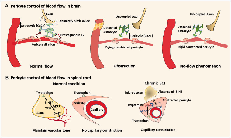
Figure 3.
Role of pericytes in blood flow control under normal and pathophysiological conditions: (a) Normally, neurons may first send signals (glutamate and nitric oxide) to astrocytes to increase astrocytic [Ca2+], triggering the release of prostaglandin E2 to relax pericytes. When obstruction occurs in pre-capillary arterioles, the low ATP increases the intercellular [Ca2+] of distant capillary pericytes, resulting in irreversible pericyte constriction even after restoration of blood flow. (b) In normal spinal cord, the vessel constrictor 5-HT is released by axons via enzymes TPH and ADCC to maintain the basal vascular tone. Since TPH and ADCC are minimally expressed by capillary cells, there is no capillary constriction with tryptophan treatment. In chronic SCI, the compromised axon loss the ability to generate 5-HT. Blood flow is controlled by pericytes where ADCC is highly expressed. Within the pericyte, tryptamine converted from tryptophan by ADCC activates 5-HT1B that causes pericyte to contract and capillary constriction.

