
FIGURE 1.
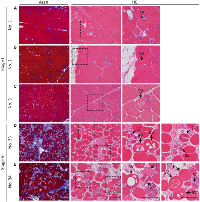
Histology of the pectoralis major muscle of broilers stained with Azan and hematoxylin–eosin. (A–E) Histological observations of the pectoralis major muscle of (A) sample number 1 (stage I; FA, 7.24% and CM, 0.70), (B) 2 (stage I; FA, 9.06% and CM, 0.69), (C) 3 (stage I; FA, 13.25% and CM, 0.68), (D) 33 (stage VI; FA, 40.90% and CM, 0.70), and (E) 34 (stage VI; FA, 40.97% and CM, 0.76). The squares indicated by dashed lines are magnified on the right. White bar = 200 mm, black bar = 100 mm. SV, myofiber with numerous small vacuoles; RV, myofiber with large-rimmed vacuoles; H, hypertrophy of myofiber; SC, myofiber with small caliber; SF, split fiber; D, myofiber degeneration; FA, fibrotic area in the muscle; CM, circularity of myofibers.