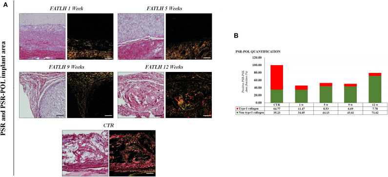
Figure 5.
Histochemical analysis of FATLH grafted in vivo using Picrosirius red staining method with white and polarized light. (A) Analysis of controls and animals grafted with FATLH at different periods of time. Scale bar = 100 μm. (B) Quantification of different types of collagen fibrers using the PSR-POL method. For each sample, the total percentage of area occupied by type-I collagen (in red) and other collagen types (in green) is shown. Values are normalized regarding the controls.

