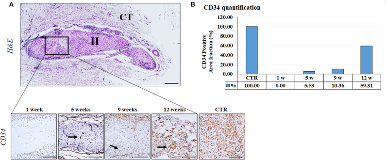
Figure 6.
CD34 Immunohistochemical analysis of implanted FATLH over the time. (A) Low magnification section of the grafted biomaterial stained with H&E (top panel). H: FATLH, CT: surrounding connective tissue. Lower panel: Positive immunohistochemical reaction for CD34 (black arrows). Scale bar = 200 μm (H&E) and 100 μm (CD34). (B) Percentage of tissue surface occupied by CD34- positive blood vessels, normalized regarding the controls.

