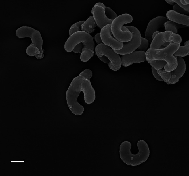
Fig. 2.

Scanning electron micrograph of c-shaped cells of Aurantimicrobium minutum KNCT. Cells were cultured in organic NSY (nutrient broth, soytone, and yeast extract; Hahn et al., 2004) medium for two weeks. Scale bar: 200 nm. This micrograph is an unpublished figure from the author; other micrographs of this species are shown in Nakai et al. (2013, 2015).
