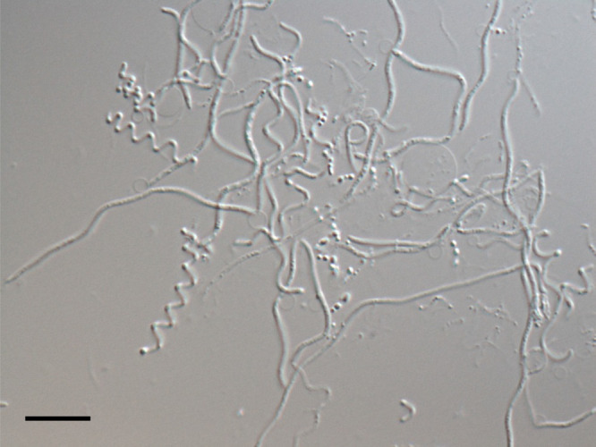
Fig. 3.

Micrograph of pleomorphic cells of Oligoflexus tunisiensis Shr3T. Cells were cultured in R2A medium for more than two weeks. This micrograph is slightly modified from the figure originally published in Nakai and Naganuma (2015). Scale bar: 10 μm.
