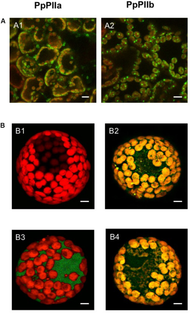
FIGURE 2.

Subcellular localization of PpPIIa and PpPIIb. (A) Localization in N. benthamiana leaves. (A1,A2) GFP-chlorophyll merged images showing respective localizations in the chloroplast of PpPIIa and PpPIIb. (B) Localization in isolated pine protoplasts. A single cell is shown containing plastids. (B1) control with no construct; (B2) GFP–PpPIIa construct containing transit peptide (cTP); (B3) GFP-PpPIIa construct without cTP; (B4) GFP-control construct without the PpPIIa protein but containing cTP. Scale bar represents 10 μm.
