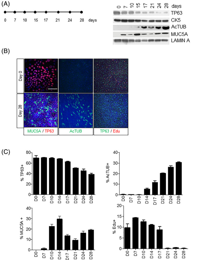
FIGURE 1.

Proliferation and differentiation of human PBECs in ALI culture. A, Western blot of the time‐dependent expression (days) of basal stem cell markers (TP63 and CK5) and differentiation markers Ac‐TUB (ciliated cells) and MUC5A (mucous cells) after airlift in ALI culture. Lamin A was used as loading control. B, Immunofluorescent costaining of PBECs at day 0 and day 28 for TP63, MUC5A; Ac‐TUB, and proliferation with EdU. C, Quantification of TP63+, Ac‐TUB+, MUC5A+ and EdU+ cells in ALI system. For each staining condition, we randomly selected five different fields. The cells in these five fields were then counted to obtain a total of 500‐1000 cells per condition (100‐200 cells per image). Stainings with TP63, Ac‐TUB, MUC5, and EdU were captured using a ×20 objective. The Z‐stack was used as the image in the paper. Image‐J was used to count the positive cells and the foci in the nucleus. Comparable results were obtained in at least three independent donors. All scale bars are 1 μm. Ac‐TUB, Acetylated Tubulin; ALI, air‐liquid interface; PBECs, primary human bronchial epithelial cells
