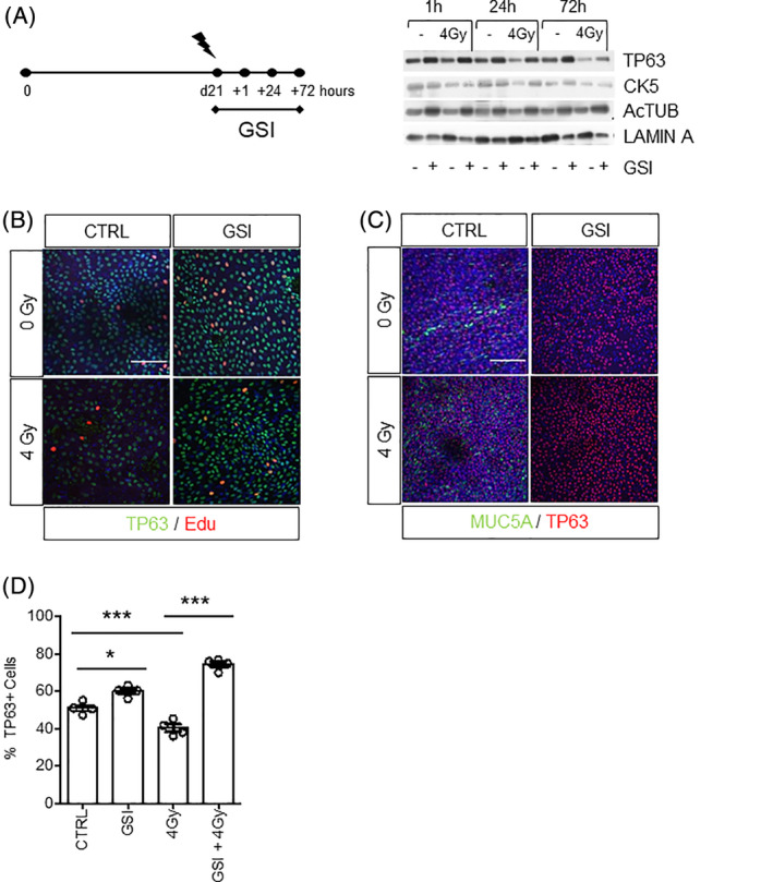
FIGURE 3.

NOTCH inhibition prevents the loss of TP63+ basal stem cells after radiation. A, Expression of TP63, CK5, Ac‐TUB 1 hour, 24 hours, and 3 days after RT of PBECs upon continuous vehicle or GSI treatment by Western blot. Lamin A was used as loading control. B, Immunofluorescent costaining of TP63 and EdU showed increased proliferation of TP63+ basal cells 3 days after irradiation upon GSI treatment. C, Immunofluorescent costaining of TP63 and MUC5A in control and GSI‐treated samples with and without irradiation. D, Quantification graph of the percentage of TP63+ cells in irradiated samples upon GSI treatment. N = 3 biological repeats per donor. One‐way ANOVA: *P < .05; ***P < .0001. All scale bars are 1 μm. Ac‐TUB, Acetylated Tubulin; ANOVA, analysis of variance; EdU, 5‐ethynyl‐2‐deoxyuridine; GSI, γ‐secretase inhibitor; PBECs, primary human bronchial epithelial cells
