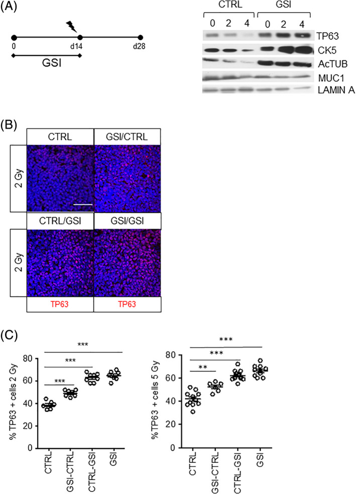
FIGURE 4.

NOTCH inhibition increases the proliferation of irradiated TP63+ cells. A, PBECs were grown in ALI in the presence of vehicle or GSI for 14 days prior to 2‐ or 4‐Gy irradiation at day 14. Cell collection was at 28 days after airlift. No GSI or vehicle was added after irradiation. Protein expression of epithelial lung cell markers TP63, Ac‐TUB, and MUC1. Lamin A was used as loading control. B, Ac‐TUB showed increased percentage of TP63 when NOTCH was inhibited in primary murine stem cells after in vivo irradiation. This effect was time‐dependent. Longer incubation with NOTCH inhibitors resulted in increased percentage of TP63+ cells. C, Quantification of TP63+ cells in irradiated mice 2 and 5 Gy. N = 3 biological repeats per donor. Statistical analysis was one‐way ANOVA: **P < .001; ***P < .0001. All scale bars are 1 μm. Ac‐TUB, Acetylated Tubulin; ALI, air‐liquid interface; ANOVA, analysis of variance; GSI, γ‐secretase inhibitor; PBECs, primary human bronchial epithelial cells
