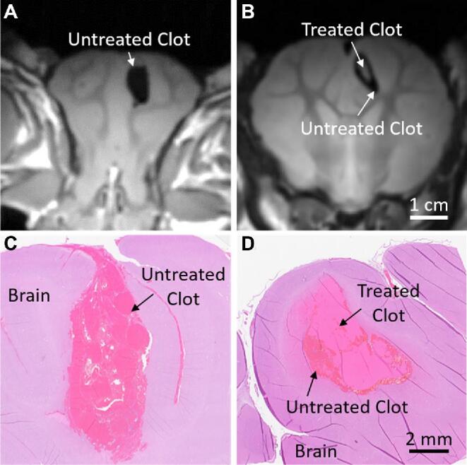
Figure 3.

The T2-weighted FSE MRI of Ex Vivo, formalin-fixed acute clot and brain for the A, untreated pig and B, treated pig. H&E-stained sections of the acute clot and brain for the C, untreated pig, and D, treated pig. Sections from the untreated pigs showed fully intact coagulated clots, whereas the clots treated with histotripsy were characterized by an acellular, homogenized core surrounded by an intact rim of untreated clot.
