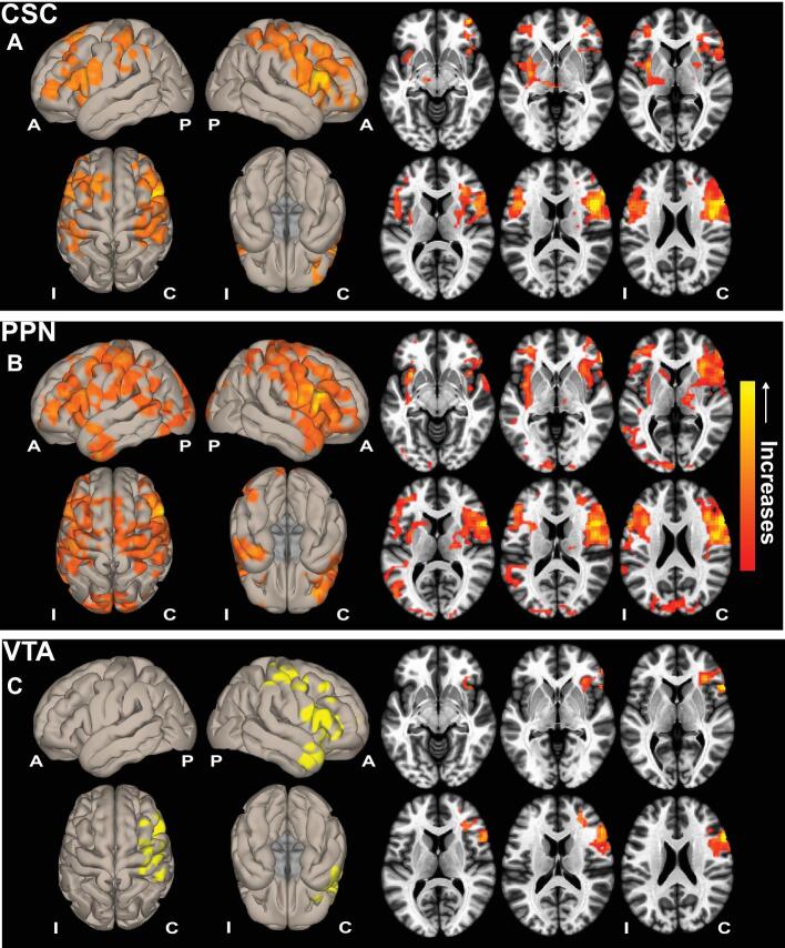
Figure 1.
ARAS functional connectivity increases in seizure-free TLE patients after surgery. Cortical surface (left) and axial slice (right) views are shown, demonstrating functional connectivity increases in patients with TLE who achieved seizure freedom after surgery, seeded from CSC A, PPN B, and VTA C. Data represent seed-to-voxel functional connectivity (bivariate correlation) maps comparing postoperative vs preoperative fMRI (paired t-test, cluster threshold level P < .05, FDR correction) generated using the CONN toolbox (https://www.nitrc.org/projects/conn/). Positive contrasts are shown, and no connectivity decreases were observed on negative contrasts. Images are oriented across all patients with respect to the epileptogenic side. N = 10 patients before surgery and > 1 yr after surgery. A, anterior; ARAS, ascending reticular activating system; C, contralateral; CSC, cuneiform/subcuneiform nuclei; FDR, false discovery rate; I, ipsilateral; P, posterior; PPN, pedunculopontine nucleus; VTA, ventral tegmental area.

