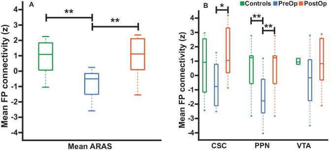
Figure 2.
ARAS-frontoparietal functional connectivity in TLE patients before and after surgery and controls. A, Mean functional connectivity between ARAS and frontoparietal and insular neocortex is reduced in preoperative patients with TLE compared to controls. However, connectivity in the same TLE patients is increased > 1 yr after surgery, resembling connectivity in controls. B, Examining ARAS regions individually, increases in frontoparietal connectivity are seen after surgery in CSC and PPN, but not VTA. n = 10 patients before surgery and > 1 yr after surgery, who ultimately achieved seizure freedom vs 10 matched controls. *P = .05, Kruskal–Wallis with post hoc Dunn; **P value range = .01-.04, Kruskal–Wallis with post hoc Dunn. Central bar shows median, bottom and top edges of box indicate 25th and 75th percentiles, and whiskers indicate data extremes. ARAS, ascending reticular activating system; CSC, cuneiform/subcuneiform nuclei; FP, frontoparietal; PostOp, postoperative patients; PPN, pedunculopontine nucleus; PreOp, preoperative patients; VTA, ventral tegmental area.

