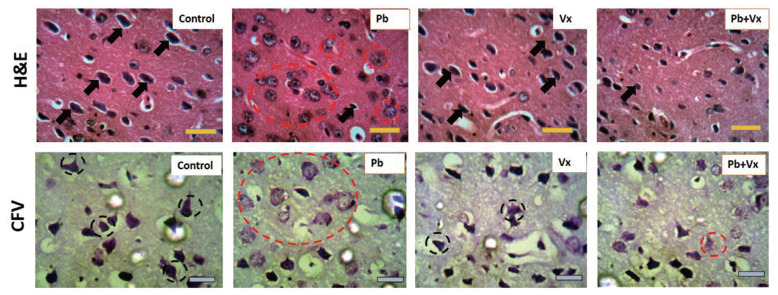
Figure 4.
Representative photomicrograph of haematoxylin and eosin (H&E) stain as well as cresyl fast violet (CFV) stain of the dorsolateral prefrontal cortex (DLPFC) in mice. Control= control group; Pb= Lead acetate treated group; Vx=Vitexin treated group; and Pb+Vx= Lead acetate + Vitexin treated group. Black arrows= pyramidal neurons; Dotted red circles= degenerative changes; Black dotted circle= intensely stained Nissl substances, Scale bar= 45 μm.

