
FIGURE 5.
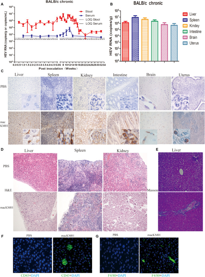
Profiles of gt4 HEV-persistent infection in BALB/c mice. HEV RNA was detected in the stool and serum (A) and tissues (B) of BALB/c mice inoculated with the stool supernatant from a chronic infected rhesus macaque (macKM01). LOQ, limit of quantification. HEV antigen was detected in the liver, spleen, kidneys, intestines, brain, and uterus of BALB/c mice by immunohistochemical method (×200; C) in BALB/c mice inoculated with macKM01, ×200. Histopathological analysis was performed in the liver, spleen, and kidneys of HEV persistent infected BALB/c mice (H&E), ×200 (D). Liver fibrosis was evaluated by Masson staining (E), ×200. Immunofluorescent staining for leukocyte (CD45+, green, F) or macrophages (F4/80+, green, G) was performed in the liver of uninfected (PBS) or macKM01-infected BALB/c mice, ×400. LOQ of stool is 2.8 × 10³ copies/g, LOQ of serum is 1.8 × 10³ copies/ml, and LOQ of tissues is 2.0 × 10³ copies/g.