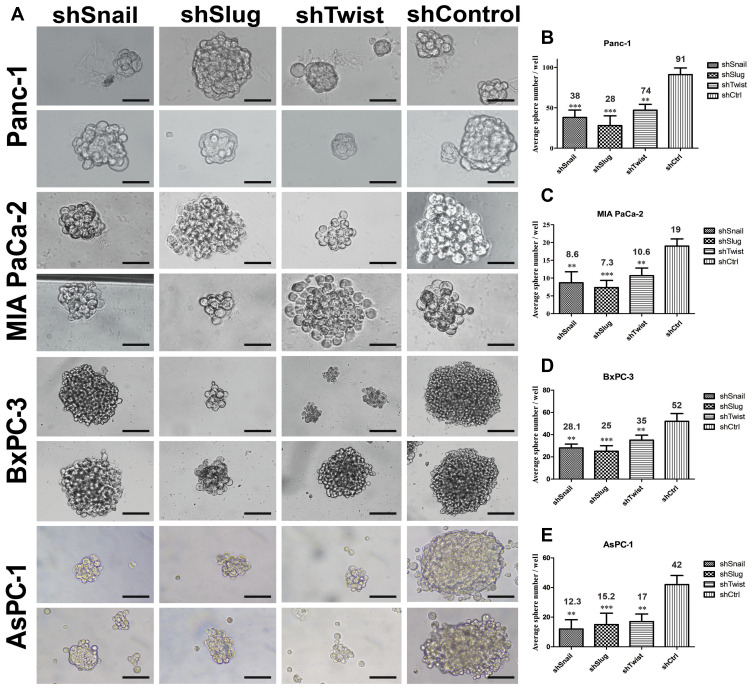
Figure 6.
EMT-ATF silencing decreased the number and the size of tumor spheres. (A) Tumor sphere images of PC cells with EMT-ATF silencing and their controls. Cellular images were taken under a light microscope with 200x magnification and spheres (size> 50 µm) were counted. Graphical representation of an average number of spheres of Panc-1 (B), MIA PaCa-2 (C), BxPC-3 (D) and AsPC-1 (E) cells with gene silencing compared to their control (**p<0.01 and ***p<0.001). The graph indicates the differences in average sphere numbers per well at 200× magnification. The values represent the mean ± SD of triplicate samples from a single representative experiment (n=9). Scale bar represents 100 µm.

