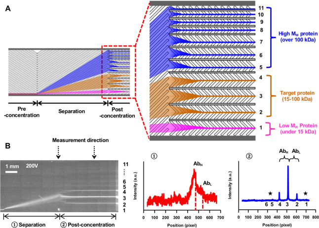
Figure 2.
Protein separation and concentration in the nanofluidic device. (A) Concentration of separated proteins in 11 individual small channels in the postconcentration region. (B) Offline size separation of commercial IgG1 (100 μg mL–1) in the nanofluidic device. The fluorescence image of IgG1 in the nanofluidic device and signal profiles in the separation and postconcentration regions. AbH, AbL, and star symbols represent antibody heavy chain, light chain, and impurities, respectively.

