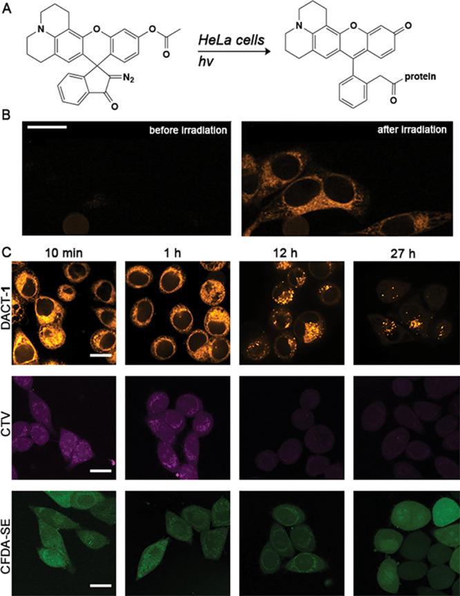
Figure 2.

Fluorescent signal durability of cells stained with DACT-1, CTV, and CFDA-SE with varying incubation times after staining protocol. (A) Schematic representation of the intracellular reaction of DACTs. (B) Confocal images obtained from treating HeLa cells with DACT-1 (10 μM) for 10 min. Photoactivation and read-out were achieved using 405 nm (1 s, 30 mW) and 561 nm lasers (0.5 s, 120 mW), respectively. (C) Comparison of fluorescence images of cells incubated with DACT-1 (10 μM for 30 min and photoirradiated), CTV, or CFDA-SE (in a solution of 5 μM dye in FluoroBright, for 30 min) individually, washed and imaged after 10 min and 1, 12, and 27 h using confocal microscopy. Representative images from three independent experiments are displayed. Scale bars = 10 μm.
