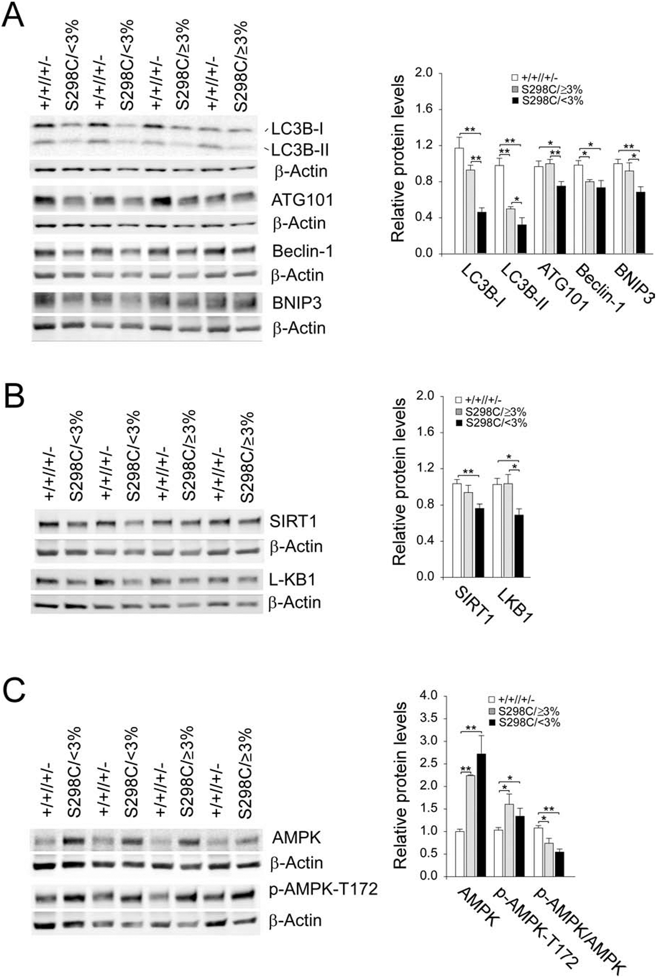
Fig. 4.

Impaired hepatic autophagy in the S298C/<3% mice. The data were analyzed from wild-type (+/+//+/− , n = 15), S298C/≥3% (n = 9) and S298C/<3% (n = 8) mice. (A) Western-blot and densitometry analyses of hepatic LC3B, ATG101, Beclin-1, BNIP3, and β-actin. (B) Western-blot and densitometry analyses of hepatic SIRT1, LKB1, and β-actin. (C) Western-blot and densitometry analyses of hepatic AMPK, p-AMPK-T172, and β-actin. Data represent the mean ± SEM. *P < 0.05, **P < 0.005.
