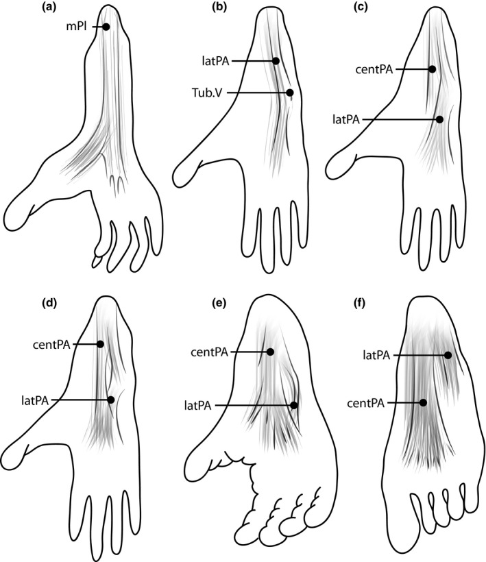
Figure 3.

Illustrations of the anatomical variation of the plantar aponeurosis among primates. (a) The plantar aponeurosis is primitively continuous with the plantaris muscle (mPl). This form is described, for example, in black‐and‐white ruffed lemurs (Varecia variegate). (b) The plantar aponeurosis forms a lateral band (latPA) that attaches proximally to the lateral tubercle of the calcaneus and distally to the joint capsule at the fifth tarsometatarsal joint Tub.V), as described, for example, in Venezuelan red howlers (Alouatta seniculus). (c–f) Various formations of the plantar aponeurosis, with a lateral and a central band (centPA). While the lateral band appears prominent in olive baboons [Papio anubis (c)] and guinea baboons [Papio papio (d)], the central band is more prominent in chimpanzees [Pan troglodytes (e)] and humans [Homo sapiens (f)].
