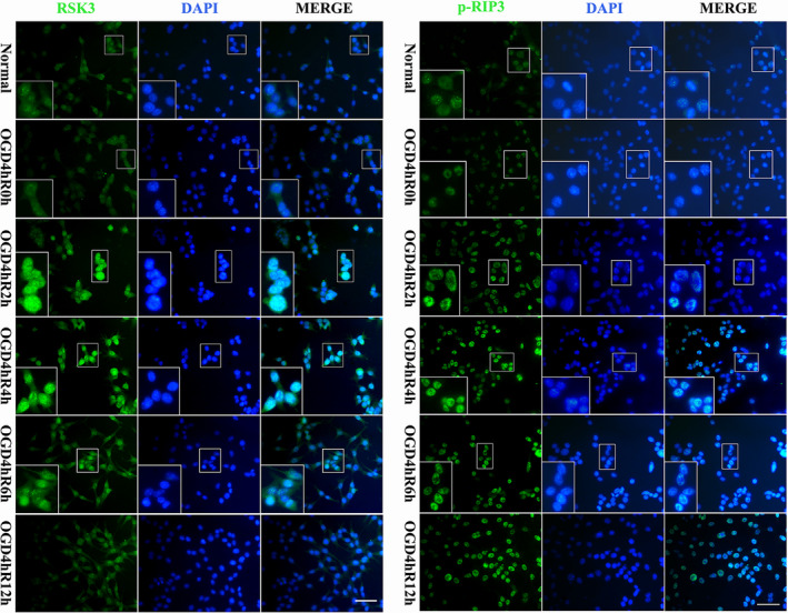
Figure 3.

Immunofluorescence staining of RSK3 and p‐RIP3 in RGC‐5 following OGD at different survival time points. Green fluorescence was positive for immunofluorescence staining of RSK3 and p‐RIP3. Blue fluorescence was the DAPI‐labeled nuclei. Scale bars: 30 μm (applies to all images except the high magnification images); 60 μm (applies to all high magnification images)
