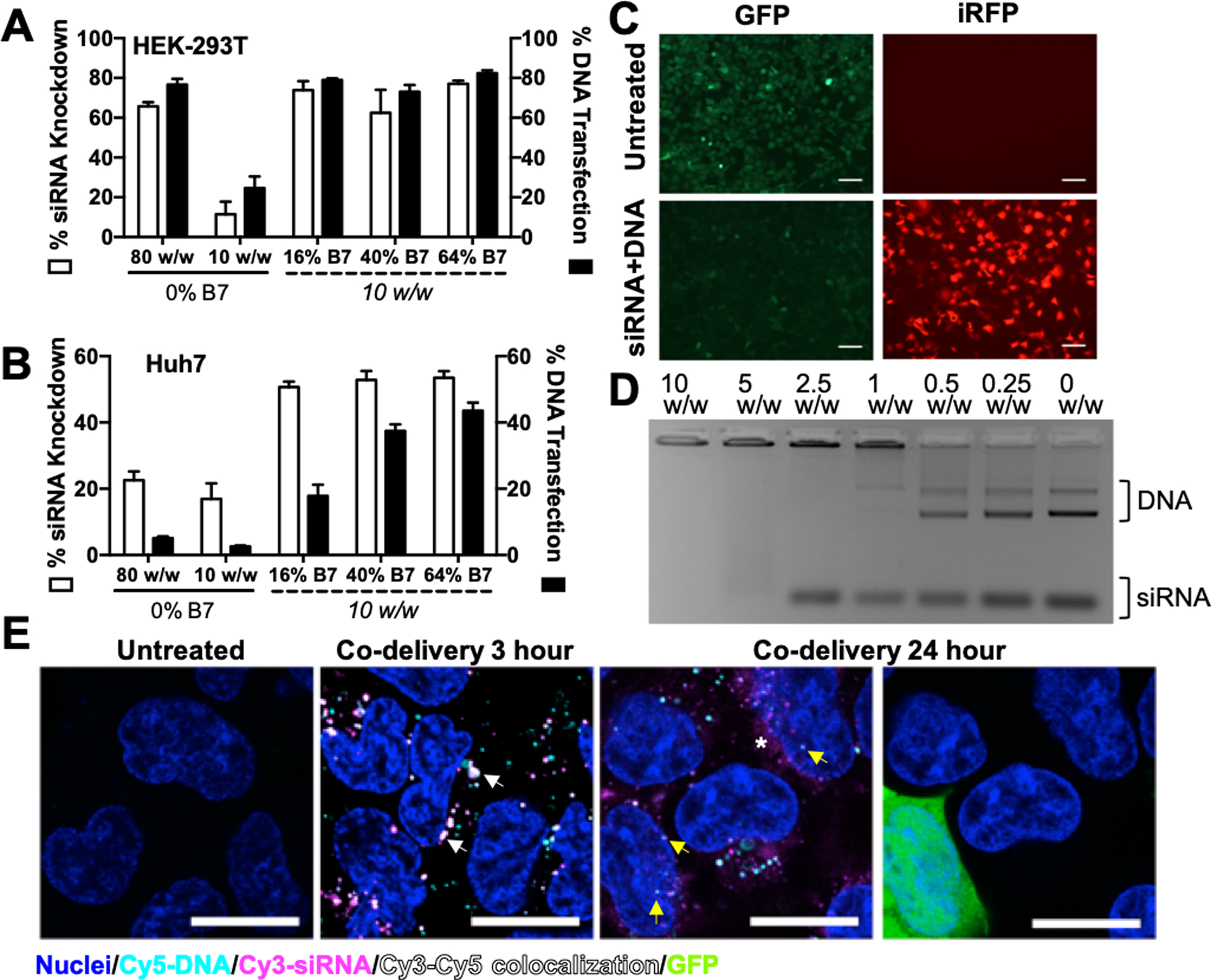
Figure 3. Hydrophobic R6,7,8-4-6 polymer series enables efficient co-delivery of DNA and siRNA.

Co-delivery efficacy of R6,8-4-6 (0% B7) and R6,7,8-4-6 nanoparticles encapsulating 400 ng total nucleic acid in 293T (A) and Huh7 (B). N = 4. (C) Fluorescence microscopy images of HEK-293T cells treated with R6,7,8_16 nanoparticles co-delivering 200 ng siRNA and 200 ng DNA (10 w/w formulation). Scale bar 100 μm. (D) R6,7,8_64 completely encapsulated plasmid DNA and siRNA at 10 w/w as seen by a gel retardation assay. (E) Confocal microscopy images of 293T cells treated with R6,7,8_64 nanoparticles co-delivering Cy3-siRNA, Cy5-DNA, and unlabeled GFP plasmid DNA (0.5:0.4:0.1 composition by weight) at 3 hr and 24 hr post-uptake. Cy3 and Cy5 signal colocalization could be seen at 3 hours post-uptake (white arrows). At 24 hours post-uptake, diffuse Cy3-siRNA signal could be seen in the cytosol (white asterisk) while some Cy5-DNA signal was detected in the nucleus (yellow arrows) and some cells were visibly expressing GFP. Scale bar 20 μm.
