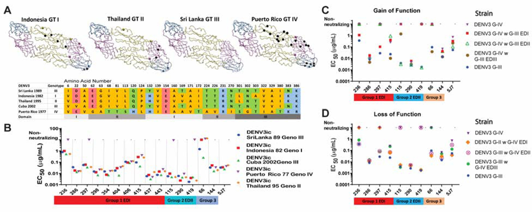
Figure 4. DENV3 genotype panel.
To identify natural variation that disrupts hmAb function, a panel of recombinant viruses were used that encoded E glycoproteins derived from the different DENV3 genotypes. Subsequently, DENV3 genotype chimeras were used to map hmAbs to EDI, EDII or EDIII in the E glycoprotein by gain-of-function or loss-of-function.
A) DENV3 E glycoprotein dimers for designated GI-IV are shown. Amino acid residues that differ from those in the Sri Lanka genotype III are shown as spheres. A black sphere indicates the residue is unique to that genotype and not shared between the genotypes. Colored spheres indicate residues seen in two or more genotypes.
B) Amino acid alignment. Amino acids with nonpolar side chains are colored orange, uncharged polar are green, acidic are red and basic are blue. Domains are indicated in gray bar at bottom.
C) Genotypic variation alters FRNT neutralization EC50 values for select DENV3 hmAbs. Genotype IV DENV3 escaped neutralization by some DENV3 hmAbs.
D) Gain-of-function genotype IV DENV3 chimeric viruses with EDI, EDII, or EDIII from genotype III DENV3 were used to map select hmAbs to specific domains of E glycoprotein (see Figure S5). EC50 values of Vero-81 cell FRNT for select hmAbs show gain-of-function for DENV-236, -297 and −415 when EDI is transplanted. DENV-115 and −419 show gain-of-function when EDII is transplanted. DENV-66 shows gain-of-function when EDIII is transplanted.
E) Loss-of-function genotype III DENV3 chimeric viruses with EDI, EDII, or EDIII from genotype IIV DENV3. EC50 values of Vero-81 cell FRNT for select hmAbs. DENV-236, −297 and −415 show loss-of-function when EDI is transplanted. DENV-115 and −419 show loss-of-function when EDII is transplanted. DENV-66 shows loss-of-function when EDIII is transplanted.

