Figure 3.
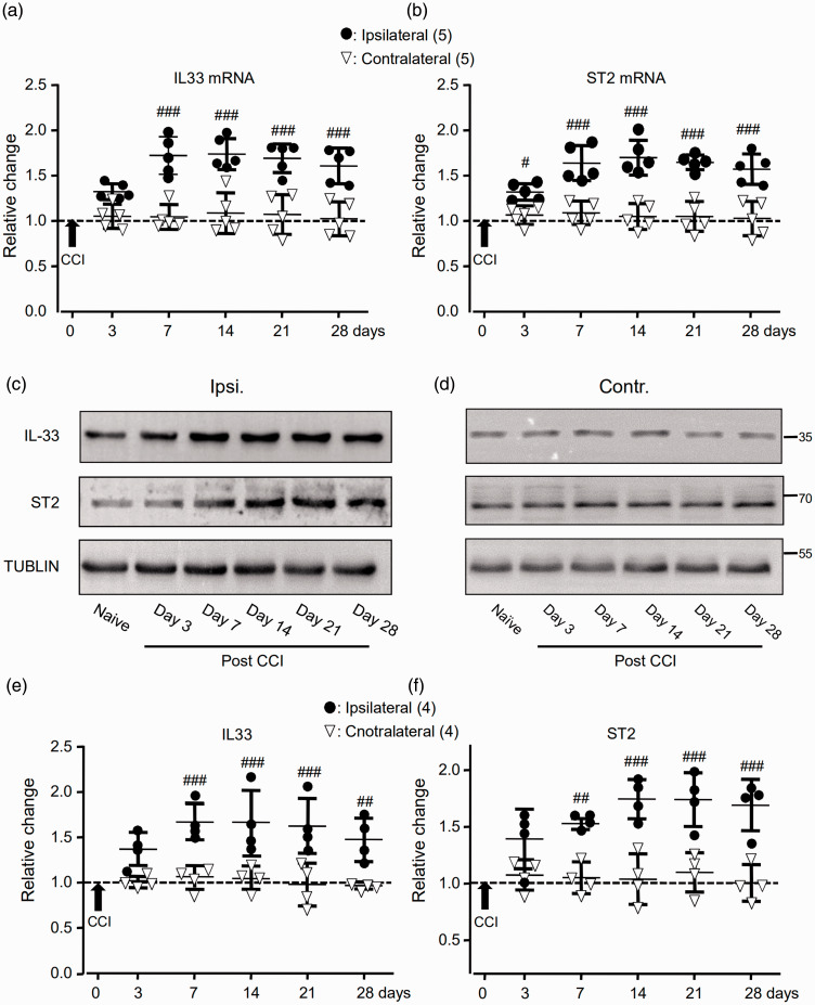
The expressions of IL-33 (IL33) and ST2 mRNAs and proteins in the SDH were increased following the CCI. (a) and (b) Summary data (mean ± SD) showing the relative changes in the expression of IL-33 (a) and ST2 (b) mRNAs after the CCI to that in naive rats (dashed line, = 1). #p < 0.05, ###p < 0.001, Bonferroni’s post hoc test in two-way ANOVA of ipsilateral versus contralateral SDH. (c) and (d) Examples of Western blots. Gels (from left to right) in (c) and (d) were respectively loaded with lysates prepared from the right L4 to L6 SDH of naive rats without any treatment (naive) or ipsilateral (ipsi.) and contralateral (contr.) SDH of rats on days 3, 7, 14, 21, and 28 after the CCI. Each group of the blots was stripped and successively probed with antibodies as indicated on the left of blots in (c). Values on the right side of blots in (d) indicate the molecular mass (Kd). (e) and (f) Summary data (mean ± SD) showing the relative changes in the expression of IL-33 (e) and ST2 (f) proteins in the SDH. The ratio of band intensities versus that of tublin was calculated and then normalized to the ratio in naive animals (= 1, dashed line) for determining relative changes. ##p < 0.01, ###p < 0.001; Bonferroni’s post hoc test in two-way ANOVA of ipsilateral versus contralateral SDH of rats after the CCI.
IL33: interleukin-33; ST2: suppressor of tumorigenicity 2; miRNA: microRNA; CCI: constriction nerve injury.

