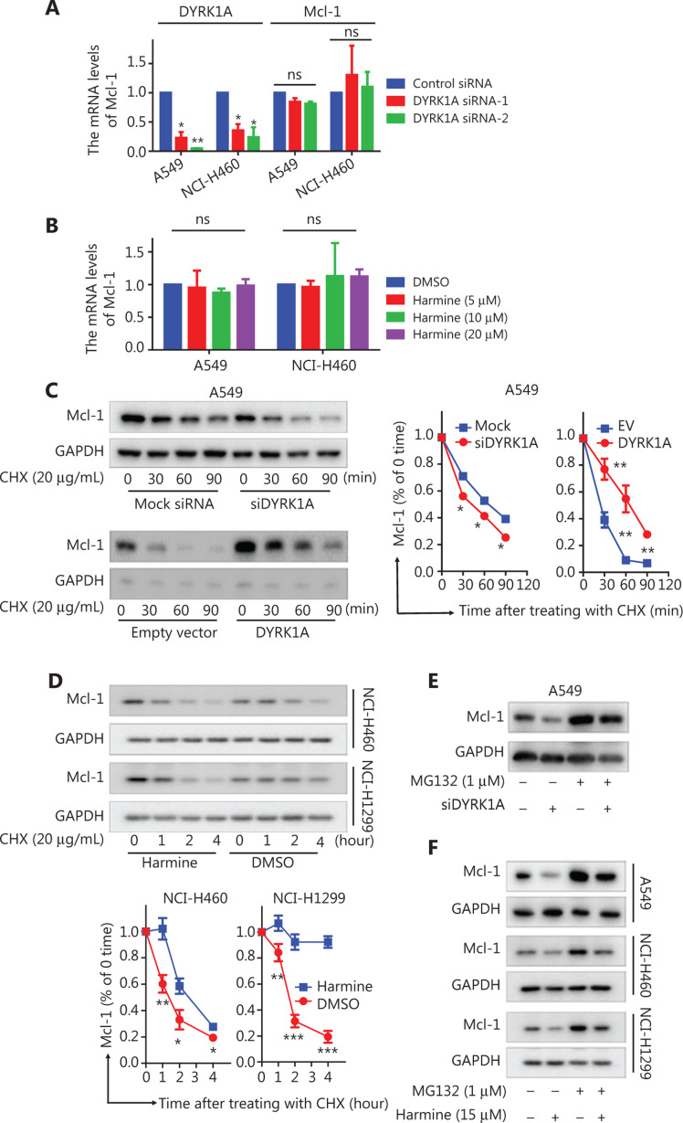
Figure 2.
DYRK1A promotes the stability of Mcl-1 in NSCLC cells. (A) NSCLC cells were transfected with control siRNA and DYRK1A siRNA for 48 h, and real-time reverse transcriptase PCR analyses were used to detect the mRNA levels of DYRK1A and Mcl-1. (B) NSCLC cells were treated with harmine for 24 h, and real-time reverse transcriptase PCR analyses were used to detect the mRNA levels of Mcl-1. (C) A549 cells were transfected with siDYRK1A or DYRK1A plasmid for 48 h. Then, cells were treated with cycloheximide (CHX; 20 μg/mL) to block new protein synthesis, and the degradation of Mcl-1 was detected by Western blot. (D) NSCLC cells were treated with CHX (20 μg/mL) in the absence or presence of 15 μM harmine for 1, 2, 3, and 4 h, after which Western blot analysis was performed to measure Mcl-1 levels. (E) A549 cells were transfected with siDYRK1A or mock siRNA for 48 h, and cells were exposed to MG132 (1 μM) or dimethylsulfoxide (DMSO) for 24 h, then Western blot analysis was performed to measure Mcl-1 levels. (F) NSCLC cells were treated with MG132 (1 μM) and/or harmine (15 μM) for 24 h, then Western blot analysis was performed to measure Mcl-1 levels. Significant results are presented as ns non-significant, * P < 0.05, ** P < 0.01, or *** P < 0.001.

